This is an automatically translated article.
The article is advised by Resident Doctor, Master Nguyen Van Anh - Radiologist - Department of Diagnostic Imaging and Nuclear Medicine - Vinmec Times City International Hospital.In the field of cancer diagnosis, especially breast cancer, one of the most commonly used diagnostic support diagnostic methods is core needle biopsy. Because it is less invasive than the traditional biopsy method but still achieves high efficiency, core biopsy is very often indicated in the diagnosis of breast cancer in recent times.
1. What is a core needle biopsy?
In order to accurately diagnose cancer diseases, including breast cancer, histopathological results through tumor biopsy are one of the indispensable criteria. Traditionally, biopsy surgery is considered the first choice, but the disadvantage of this method is that it is invasive in the patient's body and can leave some complications after surgery. Therefore, the birth of the needle core biopsy method has opened up many new directions for this field. Although less invasive and complicated than the surgical method, the effectiveness of core biopsy is on par with the old method, so it is very preferred by treating doctors during the procedure. examination and diagnosis of cancer pathology.Core biopsy is usually indicated after the patient has performed other laboratory techniques such as mammography in breast cancer. Similar to surgery, a core biopsy also allows a sample of cells to be removed from a patient's suspected tumor area, and then tested to check if the cells in that area are malignant. a cancer cell or not.
The biggest difference compared to the surgical biopsy method is that for a core needle biopsy, the patient only needs to be given local anesthesia and a needle with a core is inserted through the skin to the suspected tumor site. Meanwhile, if surgery is performed, the patient needs to undergo general anesthesia, make multiple incisions in the skin to gain access to the cancerous area and potentially leave scars after the procedure. implementation process.
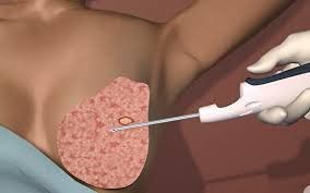
In fact, many studies have shown that the rate of complications occurring after performing core needle biopsy is very low and rarely occurs, so this is considered one of the safe diagnostic options for patients with to the present time. In general, core needle biopsies are performed using a hollow-core needle that is inserted by the physician into the patient's breast tissue after a breast ultrasound has been performed to locate a suspected tumor. Next, tissue samples will be taken and taken out through the needle core and sent to the laboratory for analysis.
Nhiều nghiên cứu đã cho thấy tỷ lệ biến chứng xảy ra sau khi thực hiện sinh thiết lõi kim là rất thấp và hiếm khi xảy ra.
2. Steps to conduct core biopsy
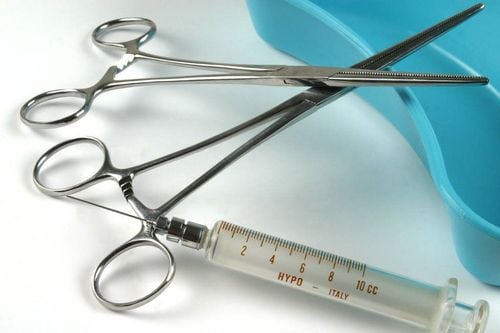
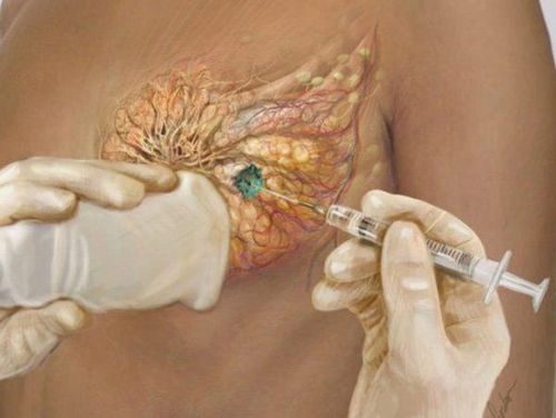
Specifically, the steps to perform the core biopsy technique are:Disinfect the skin where the needle will be inserted. Spread out the hole Anesthetize with 2ml of Novocain 3% solution. Left hand holds the ultrasound probe, and the surgeon's right hand injects anesthetic into the skin to be performed the procedure. Incision of the skin with a knife 2mm below the anesthetic site Use a Trucut to pierce the incision site so that the tip of the needle is parallel to the longitudinal axis of the ultrasound transducer and the chest wall. If the tumor is small, just insert the needle into the area about 2-3cm from the edge of the tumor 1 or right into the tumor edge for larger tumors. Perform sampling in 5 locations in 5 different directions around the tumor End the procedure by withdrawing the needle and pressing the gauze on the site for about 5 - 10 minutes. Some notes in patient care after core biopsy are:
Need for the patient to take antibiotics and pain relievers for the first 5 days. Do not perform heavy work, carry with your arm on the side of the affected breast. If you have unusual symptoms such as breast swelling, pain, and pain, you should immediately report to your doctor for treatment. If there is bruising at the needle puncture site, do not be too worried because it will automatically disappear after about 5-7 days, if it is beyond this time, notify the treating doctor. Do not shower after the procedure. Change the dressing of the puncture site regularly for the first 3 days, then remove the dressing. Specific indications for the core needle biopsy method are:
FNA aspiration has signs of cancer. Clinically there are clinical signs that suggest cancer. Ultrasound results show signs of cancer. X-ray mammogram with images of suspected breast cancer Indicated when it is necessary to differentiate between 2 types of fibroadenomas and phyllodes tumors The patient is already in stage IV cancer and his health condition does not allow surgery. Therefore, it is necessary to perform a core biopsy to find the histopathological properties of the tumor, from which to have the appropriate chemotherapy treatment for the patient. Absolute contraindications to core needle biopsies in the diagnosis of breast cancer are:
Patient is having breast prosthetic implantation Patient is experiencing clinically evident infection Patient has symptoms of: hematoma after trauma The patient has diseases related to blood clotting disorders.
Có một số trường hợp chống chỉ định tuyệt đối của sinh thiết lõi kim.
3. Conclusion
Needle core biopsy is the least complicated and invasive method of taking tissue samples for testing today, and gives very high accuracy results. Therefore, when there are any signs of doubt, the patient should immediately go to reputable and specialized medical facilities in this field for advice and guidance on the implementation of modern techniques and methods. This is very effective.Vinmec International General Hospital is applying needle core biopsy technique under ultrasound guidance to diagnose, evaluate, and detect breast cancer early.
The ultrasound equipment system at Vinmec is modernly and synchronously invested, using leading advanced equipment, making the biopsy process easier and more accurate, minimizing the risk of complications. complications after biopsy.
The needle core biopsy technique under ultrasound guidance at Vinmec is performed by a team of highly qualified and well-trained radiologists at home and abroad. Experience and expertise are the two leading factors in achieving high results with ultrasound-guided breast biopsy techniques. Therefore, patients can be completely assured when performing this technique at Vinmec.
Any questions that need to be answered by a specialist doctor as well as customers wishing to be examined and treated at Vinmec International General Hospital, please register for an online examination on the Website for the best service.
Please dial HOTLINE for more information or register for an appointment HERE. Download MyVinmec app to make appointments faster and to manage your bookings easily.