This is an automatically translated article.
X-ray-guided breast biopsy is a technique that uses a large-sized biopsy needle attached to a suction machine, using an X-ray machine as a guide to guide the needle into the correct position in order to remove the patient arrays for examination. test for early detection of breast cancer.1. What is an X-ray guided breast biopsy?
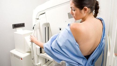
Modern digital mammography machine has the ability to position 3D space, helping to observe breast lesions clearly and accurately from different imaging directions.The doctor will proceed to select the lesion location, the machine's automatic biopsy software will determine the 3-dimensional space of that location, move the biopsy groove to the appropriate position so that it corresponds to the length. of the biopsy needle; The doctor places the needle in the biopsy groove and then pushes the needle to its full length. At that time, the needle will reach the biopsy site; The biopsy site on the skin is only about 1-2mm, completely does not affect the patient's aesthetics, even leaving no scars.
2. Indications and contraindications for X-ray-guided breast biopsy
X-ray-guided breast biopsy is indicated in cases of mammary gland lesions with high suspicion of malignancy visible only on mammography. Specifically:Suspected microcalcification lesions; Asymmetrical lesions, mass lesions, areas of structural disorder not identified on ultrasound; If there are many mammary gland lesions suspected of malignancy, biopsies of all suspected lesions should be performed to fully diagnose the lesion and assist in treatment. Contraindications to perform breast biopsy under X-ray guidance in the following cases:
People with coagulation disorders; People with serious systemic diseases such as: heart failure, kidney failure, respiratory failure; Patients with local superficial inflammation; People who are allergic to anesthetics; People are too nervous and uncooperative.

Chống chỉ định thực hiện sinh thiết vú dưới hướng dẫn X-quang trong trường hợp người đó bị suy tim
3. Equipment for performing breast biopsies under X-ray guidance
The necessary tools include:Mammogram: Used to locate the lesion; Biopsy system under X-ray, divided into 2 types: Biopsy table for prone patient: X-ray machine and biopsy system located under the table. This case helps to immobilize the patient well, reducing the risk of vagus nerve reactions during the biopsy but the cost is high, the biopsy room needs to be spacious; Biopsy system for patients in the sitting, inclined, semi-reclining position: This case helps to access the lesion site more easily and flexibly. The cost is cheap and also does not require a large space like in the case of the patient in the prone position, but has the disadvantage that it is easy to occur vagus nerve reflexes during the biopsy.
4. Procedure for performing breast biopsies under X-ray guidance
The biopsy needle will approach the site where the lesion is thought to be most visible and the distance from the skin to the lesion is the shortest. The patient will receive breast compressions in the upright CC position or the ML tilt position. The lesion to be biopsied is within the biopsy window.The first film will locate the lesion in the x, y axis. Film taken at -15 degrees and +15 degrees. The tilt position will help determine the depth of the lesion. Based on the obtained parameters, the doctor will understand the thickness of the mammary gland parenchyma and the position of the needle, avoiding the case that the needle tip comes into contact with the detector of the X-ray machine. The appropriate distance is 4mm or more from the tip of the needle to the opposite surface of the breast.
During a breast biopsy, the biopsy needle will stay in place inside the lesion. The removed patient samples will be transferred to the tray at the back of the needle. Depending on the size of the needle, there will be a corresponding number of samples taken out. For example, with a 10 - 11G needle usually takes 12 samples, a 7 - 9G needle needs 4 samples...
Normally, patient samples are removed by rotating the biopsy needle 360 degrees. However, sampling from an area of the lesion can bring the biopsy needle trough directly to the area to be sampled. If the target lesion has calcification, it is necessary to take a scan of the specimen after the biopsy to ensure the correct biopsy of the original target lesion.
In most cases, a marker will be placed on the site of the tissue just biopsied to locate the lesion and monitor the lesion later. If biopsies of many different areas, different types of markers will be used to distinguish such as dot markers, oval markers or ribbons...
Nowadays, there are many uses. The marker is made of Titanium which is compatible with magnetic resonance imaging. Many places also have stainless steel marking tools.
After conducting the biopsy, the doctor will put pressure on the biopsy area for about 5 minutes to stop the bleeding. The incision site on the skin will be covered with adhesive tape to close. The patient will be X-rayed in both positions to re-evaluate the exact location of the sample and the position of the marker.
5. Some common problems with X-ray guided breast biopsies
In some patients with small mammary glands, after biopsy, breast compression, the thickness of the breast parenchyma may not be large enough for the biopsy needle to move to the target lesion without completely penetrating the breast. touch the detector. At that time, a number of methods can be applied such as:Change the approach to the target lesion site; Inject an additional amount of saline or local anesthetic into the mammary gland tissue to increase the thickness of the mammary parenchyma; Keep the needle tray straight in the proximal position of the target lesion; Use a breast pad. Use gauze, or bandages to thicken the mammary gland parenchyma, allowing the biopsy needle to enter easily and safely; Apply the air-gap technique. Place a second mandrel between the distal part of the detector and the breast, with the portion of the biopsy window lying in a straight line between the lesion and the two forceps. For lesions located high close to the axillary tail or close to the chest wall that are difficult to biopsies under X-ray in the prone position, the patient can be rotated towards the opening of the birth table and the arms and shoulders over the open position. , which helps to expose the mammary gland parenchyma in the back and armpit, making biopsies easier.
Lesions near the chest wall or at a high position close to the tail of the armpit may be difficult to biopsies under X-ray in the prone position. In this case, it is possible to rotate the patient toward the opening of the biopsy table and leave the arm and shoulder in the open position, thus exposing the posterior and axillary parenchyma for biopsy.
6. Why should breast biopsy under X-ray guidance at Vinmec?

Bệnh viện Đa khoa Quốc tế Vinmec đang áp dụng kỹ thuật sinh thiết vú dưới hướng dẫn X-quang vô cùng hiệu quả và linh hoạt
This technique at Vinmec is carried out according to a standard and methodical process, from the examination and preparation stage to the stage of performing the biopsy and monitoring and handling complications after the biopsy. Patients will receive comprehensive care with the highest sense of responsibility and working attitude of the medical team at Vinmec. The system of modern and leading advanced machines provides maximum support for the disease diagnosis process, bringing accurate results.
Please dial HOTLINE for more information or register for an appointment HERE. Download MyVinmec app to make appointments faster and to manage your bookings easily.


