This is an automatically translated article.
The article was professionally consulted with resident doctor Nguyen Quynh Giang - Department of Diagnostic Imaging and Nuclear Medicine - Vinmec Times City International General Hospital.Ultrasound is an indispensable imaging tool in clinical practice. This is a low-cost, easy-to-implement, and highly effective means of diagnosing many diseases? So what is 2D ultrasound?
1. What is an ultrasound?
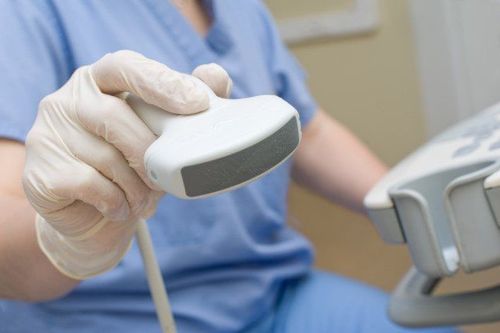
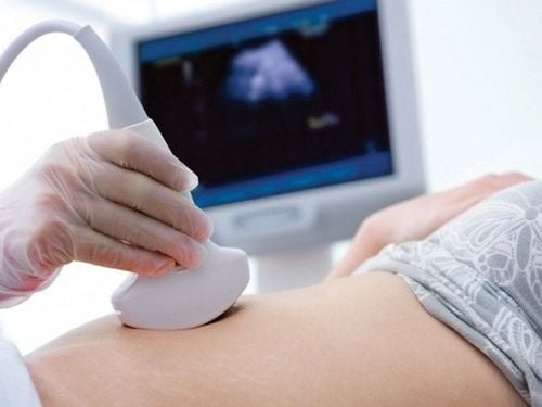
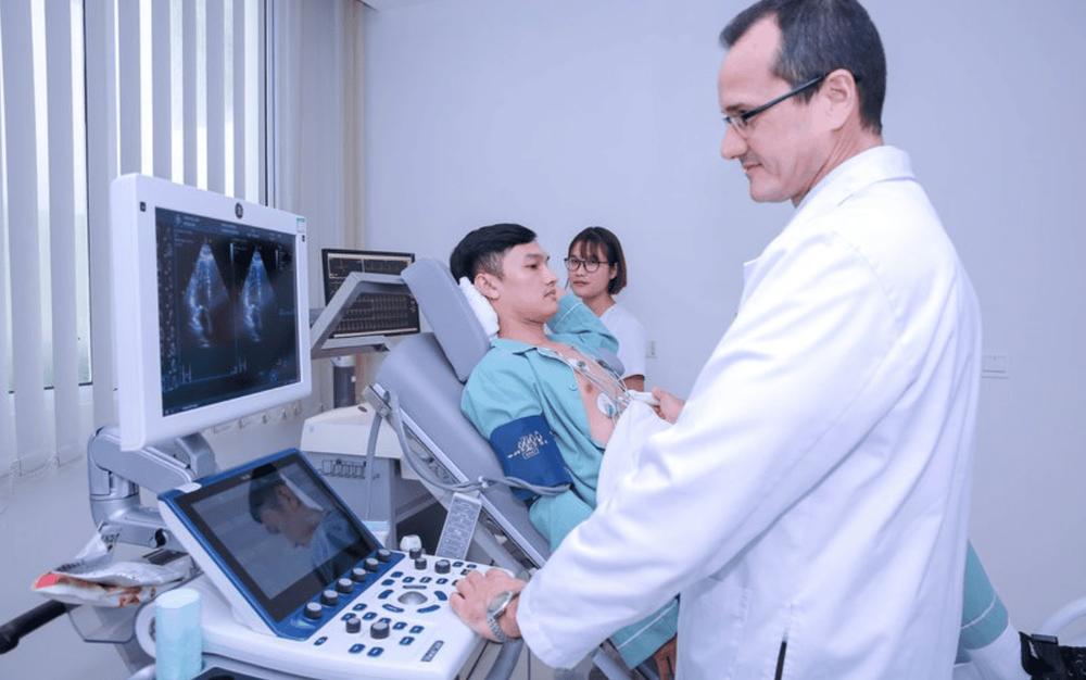
Ultrasound, English name is ultrasound - is a non-invasive technique in diagnostic imaging, used very commonly for diagnosis and treatment in medicine today in all specialties. Ultrasound technology produces structural images by rendering structures inside the body based on the tissue's high-frequency sound wave reflections - the structure, from which the sonographer will describe the features. normal or abnormal characteristics of an organization or organ in the body that cannot be observed.When doing ultrasound, the sonographer uses a transducer (transducer) close to the skin surface of the project area to be examined. The transducer has the effect of emitting high-frequency sound waves and capturing the reflected ultrasonic wave signals, then sending it to a processing and reconstruction system with organized tissue and structure to be investigated. Some have the ability to absorb or reflect sound waves to different degrees that will yield different images of the body's structures.
Ultrasound is a very popular and widely used, repeatable and routine technique without any adverse health effects.

2. What is ultrasound used for?
Ultrasound is used to examine the structures and organs of the body: Most of the components in the abdomen or abdominal cavities, the digestive system, appendix, colon, liver, spleen, pancreas, biliary tract, and gallbladder. ..., obstetrics, cardiology, urinary system, prostate gland, thyroid gland, mammary gland, musculoskeletal system, tendon attachment points, testes, uterus, ovaries... site for biopsies, intra-articular injections, vascular access and support for a number of other invasive techniques. Therapeutic ultrasound based on the effect of radiofrequency waves for some rehabilitation specialties.3. What is 2D ultrasound?
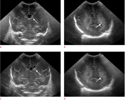
2D or 2D ultrasound: Each type A pulse wave is recorded as a bright dot more or less depending on the intensity of the echo. The movement of the transducer on the patient's skin allows to record the acoustic structure of the tissues in the body lying on the scanning plane of the sound wave beam, this is the method of ultrasound tomography. The image obtained from these echoes will be stored in memory and converted into a signal on the transmission screen by black and white and gray dots. 2D ultrasound includes:Dynamic ultrasound: It is a two-dimensional type with a fast scanning speed that creates images in real time. M-style (TM - Time Motion) ultrasound: In this type of ultrasound, the reverberation will be recorded in A-style, but moving in time thanks to the frequently panning screen. This type of ultrasound is often used for echocardiography. Doppler (Dynamic) Ultrasound: Uses the Doppler effect of ultrasound to measure circulatory velocity, determine blood flow direction, and assess blood flow. There are 3 types of Doppler: continuous Doppler, pulse Doppler, color Doppler, people often combine the Doppler system with real-time tomography ultrasound called DUPLEX ultrasound. This type of ultrasound is very convenient for cardiovascular and obstetric examinations.
4. Advantages and disadvantages of 2D . ultrasound
Advantages:Most ultrasound methods are non-invasive and usually cause no pain or discomfort to the patient. Widely used, simple and not too expensive in imaging techniques. Ultrasound does not adversely affect the body, health because it does not use radiation like techniques of X-ray, computed tomography... Ultrasound can show quite clear images of soft tissues which are often difficult to achieve. investigated by X-ray. Ultrasound can be repeated as many times as necessary for monitoring and treatment. Ultrasound provides real-time imaging results, i.e. the image changes according to the actual movements of the sonographer, making it a good tool to orient and locate for invasive procedures. such as intra-articular injections, tendon insertions, needle biopsies, and aspiration of joint or body fluids. Disadvantages of ultrasound:
Basically, ultrasound waves propagate less than in a gaseous environment, so it is difficult to survey hollow organs such as intestines, stomach and organs that are obscured by stomach and intestines such as pancreas, arteries The image in some diseases is not clearly observed by 3D, 4D ultrasound,.. Patients who are overweight, obese, and have a large waist circumference will lead to more difficult ultrasound because Fatty tissues and organs weaken the sound wave as it goes deeper into the body. Therefore, an ultrasound method cannot give the optimal image for diagnosis, the combination of other imaging techniques such as X-ray (with contrast injection), computed tomography with or without contrast. , and magnetic resonance can yield very valuable survey results. Sound waves are difficult to penetrate calcium organizations, bone tissue and therefore can only see the outside surface of bone structures, tendon attachment sites, joint sockets, but not what lies inside the bones. Bone function should be combined with other measures to determine the image of existing lesions.
Please dial HOTLINE for more information or register for an appointment HERE. Download MyVinmec app to make appointments faster and to manage your bookings easily.


