This is an automatically translated article.
This article is professionally consulted by Master, Resident Doctor Tran Duc Tuan - Department of Diagnostic Imaging - Vinmec Central Park International General Hospital.Ultrasound is a popular medical imaging technique for many years now, in which images on ultrasound play an important role to help specialists accurately diagnose conditions affecting organs in the body. body. Therefore, good imaging skills are required.
1. Ultrasound image
Ultrasound machines use transducers that emit high-frequency sound waves that affect organs and organ systems in the body to receive feedback sound waves, thereby sketching an ultrasound image. Ultrasound images are images obtained of organs and internal organs at that moment, in addition, they also show the movement and speed of blood circulation through the vascular system. Unlike x-rays, ultrasound images will not be affected by ionizing radiation.
Ultrasound images are actually created based on the reflection of sound waves when touching the organs or internal organs of the body. The intensity of this reflected sound wave combined with the time of receiving the reflection will be processed by the computer and shown through images.
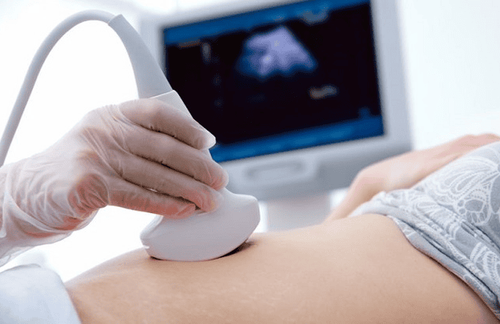
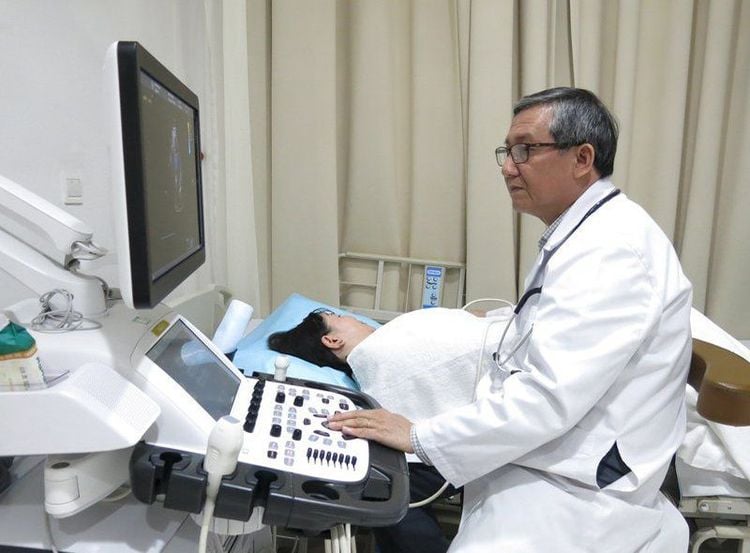
Siêu âm ghi lại hình ảnh cơ quan nội tạng trong cơ thể tại thời điểm đó
2. Ultrasonic machine structure
A basic ultrasound machine includes the following main parts:
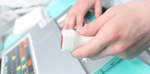
Transducer: The main part of the ultrasound machine that is responsible for and receives sound waves. The probe contains one or more voltage crystals. When an electric current is passed through these crystals, they transform, creating sound waves that propagate outward. In contrast, when receiving feedback sound waves from organs and internal organs in their body, they absorb and generate electric current for the CPU to process. Transducers come in a variety of shapes and sizes and are controlled by a probe controller shown below.
Central Processing Unit (CPU): Considered as the brain of the ultrasound machine. The CPU is basically a computer that contains the microprocessor, memory, amplifier, and power supply for the transducer. The CPU sends current to the transducer to generate sound waves later and receive electrical pulses from the transducer in return. They then perform all the calculations involved in processing the received data and output the image that appears on the screen. Transducer controller: Allows the sonographer to set and change the frequency, amplitude, time, and scan mode of ultrasound waves. Monitor : Displays an image from the data provided and processed by the CPU. There are 2 types of screens corresponding to these 2 types of ultrasound: black and white or color ultrasound Keyboard: Ultrasound machines often have built-in keyboards to facilitate data export or input as well as fill in additional notes. . Storage devices such as hard drives, floppy disks, CDs: store the acquired images. Typically, a patient's ultrasound results are stored as floppy disks in their medical records. Printer: Many ultrasound machines have a built-in printer that can print images and results from the screen.
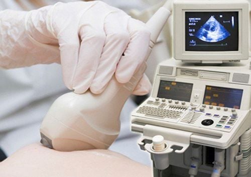
Một máy siêu âm cơ bản bao gồm 7 bộ phận chính
3. How does an ultrasound machine work?
How does the ultrasound machine work? The following are the main steps in the operation of an ultrasound machine:
1. An ultrasound machine transmits high-frequency sound waves (range 1 to 5 megahertz) into the patient's body through a transducer. 2. Sound waves transmitted into the body collide with the boundary between tissues, for example, between water and soft tissue or between soft tissue and bone. 3. Some sound waves are reflected back to the transducer while others continue to propagate until they meet boundaries between other tissues. 4. The reflected sound waves are received by the receiver, converted into electrical current and forwarded to the central processing unit (CPU). 5. At the CPU, the computer calculates the distance from the transducer to the tissue or organs through the speed of sound in the tissue (about 1540m/s) and the time to receive the reflected wave back (usually about one millionth of a second). ). 6. The machine displays the distance and intensity of the reflected wave on the screen as a 2-D image, black and white or color depending on the type of ultrasound machine.
3. Common types of ultrasound-based imaging
Ultrasound imaging is one of the important tools in diagnostic imaging, helping doctors determine the exact cause of the disease as well as the appropriate treatment.
Common types of ultrasound-based imaging diagnostics can include:
Abdominal ultrasound: Helps detect pathological conditions of some organs and internal organs in this area Bone ultrasound: Evaluation bone brittleness Breast ultrasound : Early detection of breast cancer
Siêu âm vú giúp phát hiện sớm nguy cơ bị ung thư vú
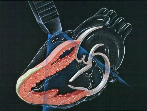
Doppler ultrasound : To visualize blood flow through blood vessels, organs or other parts of the body. Echocardiography: In order to detect early heart-related diseases such as heart valve regurgitation, heart valve stenosis... Fetal ultrasound: Helps parents monitor fetal growth, early detect abnormalities and find appropriate treatment Ophthalmic ultrasound : Determining the structure of the eye, diseases and diseases of the eye An ultrasound image is the output result of the ultrasound process from the emission of high frequency sound waves directed at enter the body and then receive reflected sound waves from tissues, organs, internal organs... through processing to produce images displayed on the screen. Ultrasound images include several types such as: echocardiogram, abdominal ultrasound, bone ultrasound, fetal ultrasound... are one of the important tools to help doctors make accurate diagnoses. disease for appropriate treatment.
Vinmec International General Hospital system uses the most modern generations of color ultrasound machines today in examining and treating patients. One of them is GE Healthcarecar's Logig E9 ultrasound machine with full options, HD resolution probes for clear images, accurate assessment of lesions. In addition, a team of experienced doctors and nurses will greatly assist in the diagnosis and early detection of abnormal signs of the body in order to provide timely treatment.
Master, Doctor Tran Duc Tuan is well-trained in the country and many centers have prestigious medical background in the world such as: Australia, Singapore, Thailand... The doctor has many years of experience in the field of medicine. imaging and endovascular and extravascular interventions. Currently, the doctor is working at the Department of Diagnostic Imaging and Interventional Vascular Unit - Vinmec Central Park International General Hospital.
To register for examination and treatment at Vinmec International General Hospital, you can contact the nationwide Vinmec Health System Hotline, or register online HERE.
References: fda.gov; webmd.com; physics.utoronto.ca; medicalnewstoday.com


