This is an automatically translated article.
This article is professionally consulted by Master, Resident Doctor Tran Duc Tuan - Department of Diagnostic Imaging - Vinmec Central Park International General Hospital.Doctors use ultrasound imaging to diagnose conditions affecting organs and soft tissues in the body, including the heart and blood vessels, liver, gallbladder, spleen, ...... , ultrasound has some limitations. So, how is ultrasound used in medical diagnosis?
1. What is an ultrasound?
Ultrasound is one of the imaging-based paraclinical methods to help doctors diagnose and monitor the patient's disease process. Ultrasound can be divided into 2 types: Diagnostic ultrasound and therapeutic ultrasound
1.1. Diagnostic ultrasound:
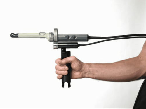
Is a diagnostic technique based on images taken and recorded in the body through ultrasound probes. These transducers produce a sound wave with a frequency much higher than the threshold of human hearing. They can be placed on the skin however to obtain the clearest and most accurate images, the transducers are placed inside the body through the digestive tract, vagina or blood vessels. This technique is also known as endoscopic ultrasound. Some basic applications of endoscopic ultrasound include:
Transesophageal echocardiography: Helps capture detailed images of the heart and major blood vessels to detect some common problems in the heart such as: valvular regurgitation, intracardiac thrombus, or aneurysm detection. Rectal transducer ultrasound: allows to evaluate the function of uterus, ovaries for women and prostate, seminal vesicles for men with high accuracy and quickly. In addition, women can also evaluate the function of the reproductive organs through transvaginal ultrasound.

Siêu âm đầu dò trực tràng giúp đánh giá chức năng của bộ phận sinh sản
Besides, transrectal ultrasound is also used during surgery by placing sterile probes into the surgical area. Diagnostic ultrasound can be divided into two types: anatomical ultrasound and functional ultrasound. Anatomical ultrasound captures images of organs, organ systems, internal organs or other structures in the body while functional ultrasound depicts the movement and speed of blood circulation, softness and stiffness of tissues or other physical features in the body. These two types of ultrasound work together to provide doctors with information about changes or differences in the function of an organ or organ system, thereby making accurate diagnoses. 1.2. Therapeutic ultrasound:
Similar to diagnostic ultrasound, therapeutic ultrasound also uses high-frequency sound waves, but the output results are not shown through images. The purpose of therapeutic ultrasound is similar to the use of physical therapy methods, which have the effect of healing wounds, heating tissues, dissolving blood clots, delivering drugs through the skin or delivering drugs to organs. specific position. In addition, ultrasound therapy is also used to destroy diseased or abnormal tissues such as tumors in cancer. The advantage of this therapy is that there is usually no need to use cutlery to perform the procedures on the body, so there are no wounds or scars.
2. Ultrasonic technical process
Before performing the ultrasound, the doctor will ask the patient to do some of the following:
Remove all jewelry that is worn on the body or at least in the area to be performed the ultrasound Take off pants or Dress in the area to be ultrasound and wear the patient's clothes Lie on the hospital bed to conduct the ultrasound

Bệnh nhân cần tháo bỏ mọi trang sức trước khi tiến hành siêu âm
During the ultrasound:
The technician will first apply an ultrasound gel to the area to be examined. This gel works to prevent the appearance of air bubbles between the transducer and the body that can affect image quality. In addition, the ultrasonic gel is not harmful and can be easily washed or washed off when it gets on clothes. The technician will then use a small hand-held device called a transducer that is gently placed and moved over the area to be ultrasounded. This transducer will emit ultrasound waves towards parts of the body and then receive feedback from the echoes to send to a computer for analysis and processing to create images. In cases where it is necessary to obtain specific images from the internal organs of the body, the probes are attached to a flexible scope and inserted through the natural openings of the body such as the mouth, anus, and vagina. ..
The ultrasound procedure usually does not cause pain to the patient, but the patient may feel a little discomfort in the case of having to insert the ultrasound probe into the body. A typical ultrasound takes anywhere from 30 minutes to 1 hour. After the ultrasound, the technician will wipe off the gel on the skin, ask the patient to wear clothes, jewelry and wait for the results according to regulations.
The patient's results will be analyzed and reported back to the treating doctor to make a diagnosis and appropriate treatment.

Bác sĩ dựa vào kết quả siêu âm để chẩn đoán về tình trạng sức khỏe người bệnh
Today, ultrasound has become more and more popular in the diagnosis and treatment of diseases because of its simplicity and accuracy. It only takes about 30 minutes to an hour and some ultrasound images can help doctors make an accurate diagnosis. In addition to being useful in diagnosing ultrasound, it can also help pregnant mothers to regularly check the development of the fetus to detect abnormalities early and to have a quick and appropriate treatment.
Vinmec International General Hospital system uses the most modern generations of color ultrasound machines today in examining and treating patients. One of them is GE Healthcarecar's Logig E9 ultrasound machine with full options, HD resolution probes for clear images, accurate assessment of lesions. In addition, a team of experienced doctors and nurses will greatly assist in the diagnosis and early detection of abnormal signs of the body in order to provide timely treatment.
Master, Doctor Tran Duc Tuan is well-trained in the country and many centers have prestigious medical background in the world such as: Australia, Singapore, Thailand... The doctor has many years of experience in the field of medicine. imaging and endovascular and extravascular interventions. Currently, the doctor is working at the Department of Diagnostic Imaging and Interventional Vascular Unit - Vinmec Central Park International General Hospital.
To register for examination and treatment at Vinmec International General Hospital, you can contact the nationwide Vinmec Health System Hotline, or register online HERE.
References: nibib.nih.gov; mayoclinic.org; radiologyinfo.org


