
This is an automatically translated article.
The article was professionally consulted with Master, Doctor Nguyen Van Huong - Department of Diagnostic Imaging - Vinmec Danang International General Hospital.An upper gastrointestinal X-ray is a form of X-ray combined with a barium contrast agent to create pictures of the inside of the esophagus, stomach, and duodenum. It is a safe, non-invasive imaging tool that can be used to help diagnose ulcers and tumors of the esophagus, stomach, and duodenum; Gastro-esophageal reflux and other upper gastrointestinal symptoms.
1. What is a gastroduodenal X-ray?
An upper gastrointestinal X-ray, also called a gastroduodenal X-ray, which sometimes also includes the esophagus, is an imaging test that uses X-rays. It is a non-invasive medical test. Invasion helps doctors diagnose and treat medical conditions based on abnormal images of structures inside the body. Radiographs are the oldest and most frequently used form of medical imaging, widely available in many places.To create clear and sharp images of the upper gastrointestinal tract, doctors often ask patients to use a special form of X-ray called TV-enhanced radiography and an oral contrast agent. orally as barite. At this time, through the television lighting, it is possible to see the internal organs in motion; whereas with barite-coated upper gastrointestinal images, radiologists can view and evaluate the anatomy and function of the esophagus, stomach, and duodenum more accurately.
2.When to take a gastroduodenal x-ray?
Gastroduodenal radiographs help examine and evaluate the structure and function of the upper gastrointestinal tract and can detect: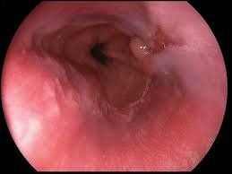
Chụp x quang dạ dày tá tràng có thể phát hiện bệnh nhân bị viêm thực quản
Tumor pushing into the lumen of the gastrointestinal tract Inflammation of the esophagus, stomach, and duodenum Indirect hernias Scars, traction, Obstruction, Structural and motility abnormalities of the gastrointestinal muscular wall Abnormalities often anatomical, such as volvulus Furthermore, gastrointestinal radiographs are also used to help diagnose the cause of symptoms such as:
Difficulty swallowing Chest and abdominal pain Gastroesophageal reflux Nausea and vomiting Unexplained persistent vomiting Indigestion Dyspepsia with blood, high suspicion of gastrointestinal bleeding
3. What do X-ray images of the stomach - duodenum show?
Gastric Ulcers The detection of gastric ulcers and their likelihood of being benign or malignant is an important goal of barium gastric radiography.Ulcers are best identified on double-contrast gastrointestinal radiographs, through signs of barite accumulation in the ulcer mouth. The most common location of peptic ulcers is in the back wall of the stomach. Meanwhile, ulcers secondary to non-steroidal anti-inflammatory drugs and alcoholism are often found on the great curvature.
In cases where folds radiating from the ulcer are observed to be smooth and symmetrical to the folds of the normal area, the ulcer can be identified as benign, usually located on the minor curvature. Conversely, if the folds converge to the site of the ulcer, incomplete healing, and signs of mucosal tissue loss are present, it should be suggestive of malignancy.
Duodenal ulcer Duodenal ulcer can also be identified by double-contrast radiographs of the duodenum and sometimes also find complications such as fistula formation with the bile duct.
Stomach stenosis Under the contrast image when taking barium, the stomach wall is markedly increased, making the stomach look like a rigid tube, which will lead to the diagnosis of gastric stenosis.
The most common cause is gastric carcinoma. The tumor invades the gastric wall, producing a thickening and fixing reaction of the gastric wall. When viewed through the magnifying screen, gastric wall peristalsis did not pass this tumor.

Polyp dạ dày
Most hyperplastic polyps have a well-defined round or oval shape, measuring less than 1cm. They have smooth contours and no evidence of contrast material within their lumen. The most concentrated site is in the muscle or body of the stomach.
In contrast, an adenomatous polyp is a true tumor because it shows a tendency to malignant transformation such as being larger than 1cm in size, lobed or tubular in shape, and especially in terms of size variation over time.
Other lesions that may also present as intraluminal masses that need to be distinguished from polyps are lymphoma, carcinoid tumor, and metastasis.
Extrinsic Lesions Lesions outside the gastroduodenal wall, forming a mass that can retract into the lumen of the gastrointestinal tract and cause external signs or obvious filling defects.
Examples of these lesions are cysts arising from the kidney, spleen, and pancreas.
4. What are the benefits and limitations of a gastroduodenal x-ray?
Benefits Upper GI radiography is a generally safe, non-invasive procedure. Indirectly evaluate the mucosa of the esophagus, stomach and duodenum. Barite is an oral contrast agent, not absorbed into the bloodstream, allergic reactions are extremely rare.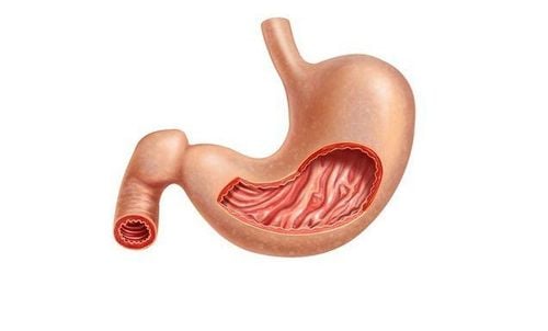
Chụp x quang dạ dày tá tràng có thể giuớ bác sĩ gián tiếp đánh giá được hình ảnh trên niêm mạc dạ dày
In addition, gastroduodenal radiographs are imaging only and cannot suggest the presence of Helicobacter pylori infection, the most common cause of ulcers. Accordingly, when it is determined that a peptic ulcer is present, the patient still has to continue to perform other invasive tests to find the cause, such as blood tests, breath tests or endoscopy, which can lead to gastrointestinal bleeding. should be combined with biopsy.
Finally, there is the risk of radiation exposure on gastrointestinal radiographs. However, given the relatively low beam intensity, the risks are insignificant compared to the benefits achieved.
In summary, before patients come to the clinic because of persistent epigastric pain, nausea and vomiting, besides upper gastrointestinal endoscopy, gastrointestinal X-ray is also a useful imaging tool. with minimal invasiveness. By administering barium or other contrast media to the patient, the gastrointestinal X-ray image becomes clearer and the diagnosis easier to confirm.
In order to improve the examination and treatment process, Vinmec International General Hospital has applied gastrointestinal X-ray technique in examination and diagnosis of many diseases. X-ray technique at Vinmec is carried out methodically and according to standard procedures by a team of highly skilled doctors and nurses, modern machinery system, thus giving accurate results, making a significant contribution to the determine disease and disease stage.
For consultation and examination at Vinmec medical system nationwide, please register at the Website for the best service.
Please dial HOTLINE for more information or register for an appointment HERE. Download MyVinmec app to make appointments faster and to manage your bookings easily.