This is an automatically translated article.
The article is professionally consulted by Master, Doctor Le Xuan Thiep - Department of Diagnostic Imaging - Vinmec Ha Long International Hospital
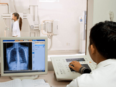
Esophageal X-ray is a classic imaging method that uses contrast dye to image the esophagus. This is a highly valuable method in locating the lesion, determining the extent of the lesion in the esophageal wall.
1. What is an esophageal X-ray?
Esophageal X-ray is the classic imaging method, which is esophagography using contrast, but still has high value in locating the lesion and determining the extent of the lesion in the wall of the esophagus. manage.General principles when performing an esophageal x-ray include:
Multiple positions: Straight, inclined, right and left obliques Kinetic study: One sip at a time Segmentation note contraindications for this patient with contrast material Special position: Hernia, reflux

X-quang thực quản là phương pháp chẩn đoán hình ảnh cổ điển là chụp thực quản có sử dụng thuốc cản quang, nhưng vẫn có giá trị cao trong việc định vị tổn thương và xác định mức độ lan rộng của tổn thương tại thành thực quản
2. Esophageal x-ray image
2.1 Anatomical X-ray of the esophagus Normal image of the normal esophagus consists of 3 segments:Cervical esophagus: Fairly short Thoracic esophagus: Located anterior to the spine with a clear margin with two concaves in the border. anterior left by the arch of the aorta and left main bronchus pressing on the abdominal esophagus: Very short and terminated by the cardia located posteriorly to the medial posterior aspect of the great gastric aneurysm and forming with this part an acute angle known as the angle of His. After each sip of barium passes, the lumen of the esophagus closes on its own and the baryte is deposited between the mucosal folds forming 2-3 regular parallel lines. 2.2 Abnormal Esophageal X-ray Anatomical Imaging Abnormal esophageal x-ray anatomical images in some symptoms are divided into two categories:
2.2.1 Esophageal dysfunction Movement disorders due to secondary or anti-peristaltic waves. These waves usually originate from the lower part of the thoracic esophagus and propagate upward, which are common in early-stage achalasia.
These signs are visible when projected on a brightening screen. For third-wave dyskinesia: along the length of the esophageal border, shallow depressions appear, which, if severe, produce multistage constriction.
2.2.2 Physical lesions Esophageal stricture: The malignant esophageal stricture is often deviated, with irregular margins, and the junction between the healthy part and the sloping part of the lesion. In the case of benign stenosis, the segment is straight axial, the border is regular, and the junction between the healthy part and the damaged part changes slowly. Defect: Formed by a proliferative mass in the lumen of the esophagus or the esophagus being pressed from the outside. This image may be located at the esophageal margin or on the surface creating small or smooth macular opacities, depending on the cause. Ulcers: These are images of drug deposits protruding above the esophageal border, usually in the lower 1/3 of the esophagus due to acid reflux Hard image: A segment of the esophageal border loses its peristaltic wave and may Intraluminal collapse represents focal thickening of the esophageal wall, common in infiltrative cancers

Hình ảnh giải phẫu thực quản bình thường gồm có 3 đoạn: thực quản cổ, thực quản ngực, thực quản bụng
3. Some common diseases in the esophagus
3.1 Esophageal cancer Esophageal cancer is diagnosed based on biopsy combined with endoscopy. Esophageal X-ray is performed before surgery or when the scope cannot pass through the stricture, and plays a role in determining the location, length, upper and lower limits of the lesion, and evaluate the condition of the esophagus. stomach condition.X-ray images in esophageal cancer include:
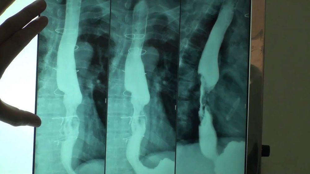
Infiltrates: Axial narrow, zigzag, crooked. If at an early stage can see hard chiseled shape. Tumor form: The image is irregular with the border, the large buds can block the esophagus. Canker sores This form of cancer presents as an ulcer surrounded by a bright rim called a lens ulcer. 3.2 Esophagitis Caustic esophagitis: Initial symptoms of oesophagitis are inflammation due to burns after narrowing due to fibrosis scars. The esophagus is usually narrow in the lower 1/3, narrow in the long axis, and has a regular border. Pass through the narrow space continuously. Radiation esophagitis: Usually occurs 6 months after radiation treatment. The narrow segment is localized in the irradiated area, sometimes accompanied by ulceration. 3.3 Esophageal Enlargement or Cardiac Constriction The esophagus is two, three to four times larger than normal. Long and curved to the right, creating an irregular opacity of the right mediastinum on chest radiographs. Baryte esophagogram shows that the esophagus is narrow at the heart, straight axis, and the esophagus is dilated evenly above.
These signs make the esophagus look like a radish, the tip of a sword. Circulation through the cardia is not continuous because it occurs only when the weight of the food and fluid exceeds a certain threshold to overcome the force of contraction in the heart. This circulation explains the absence or very little gas in the stomach.
3.4 Esophageal diverticulum: Contrast pouch with neck attached to the wall of the esophagus, usually located in the middle 1/3 or just above the diaphragm. The size of the diverticulum varies in size, can be very large, and is located just above the right border of the esophagus, above the diaphragm.
3.5 Esophageal varices In the case of portal hypertension, the submucosal venous plexus of the esophagus must ensure the return of portal blood to the vena cava. As a result, it will be stretched to create concentrated circular highlights such as clusters of grapes or long zigzag light trails, and sometimes create a fake image.
3.6 Diaphragm hernia There are many types of hernia including:
Sliding hernia: This is the most common type, the hernia slips through the diaphragmatic foramen and pulls the stomach aneurysm above the diaphragm. esophageal: the cardia is located below the diaphragm, only the air sacs pass through the diaphragm and rest on the diaphragm. Sliding and rolling hernia: Common in the elderly, can be combined in case of large hernia mass. Sliding hernia with congenital short esophagus: The esophagus is short, the air sac is located on the diaphragm. This type is often accompanied by esophagitis leading to stricture and sometimes esophageal ulceration.

Một số bệnh lý thực quản có thể gặp như là ung thư thực quản, thoát vị hoành, giãn tĩnh mạch thực quản,...
X-ray technique at Vinmec International General Hospital is conducted by a team of highly qualified and experienced medical doctors; the system of advanced and modern machinery should significantly reduce radiation dose with the best image and always be updated according to medical progress in the world; Especially, professional service quality will help customers have the most comfortable and secure experience when visiting Vinmec.
In addition, at Vinmec International General Hospital, there is a package for screening and early detection of cancers of the gastrointestinal tract (esophagus - stomach - colon) combining clinical and subclinical examination to bring results. as accurate as possible.
When screening for gastrointestinal cancer at Vinmec, you will receive:
Gastrointestinal specialty examination with an oncologist (by appointment). Gastroscopy and colonoscopy with an NBI endoscope under anesthesia. Complete peripheral blood cytology (by laser counter). Automated prothrombin time test. Automated thrombin time test. Activated Partial Thromboplastin Time (APTT) test using an automated machine. General abdominal ultrasound
Please dial HOTLINE for more information or register for an appointment HERE. Download MyVinmec app to make appointments faster and to manage your bookings easily.


