This is an automatically translated article.
The article was professionally consulted by Specialist Doctor I Tran Cong Trinh - Radiologist - Radiology Department - Vinmec Central Park International General Hospital. The doctor has many years of experience in the field of diagnostic imaging. And Specialist Doctor I Nguyen Truong Duc - Doctor of Radiology - Department of Diagnostic Imaging and Nuclear Medicine - Vinmec Times City International General Hospital.Pelvic magnetic resonance imaging (MRI) is an imaging test that uses a machine with powerful magnets and radio waves to create images of organs located in the mid-pelvic region. These structures include the bladder, prostate gland in men, ovaries and uterus in women, lymph nodes, large intestine, small intestine, and pelvis. Because it is obscured by the skeleton, magnetic resonance imaging is the ideal means for investigating the pathologies here.
1. What is a magnetic resonance imaging (MRI) of the pelvis?
Magnetic resonance imaging (MRI) is a noninvasive test used to diagnose medical conditions. An MRI uses a strong magnetic field, radio waves, and a computer to create detailed images of the body's internal structures without using radiation energy (X-rays) like X-rays or computed tomography (CT) scans. ).Detailed MRI images allow doctors to examine organs and detect abnormalities. Images can be viewed on a computer screen or can be sent electronically, printed or copied to a CD, or printed into film. Any area of the body, if there are no contraindications, can have an MRI.
The pelvic MRI area is usually performed with the abdomen, with solid and hollow organs in the abdomen, including: liver, biliary tract, kidney, spleen, intestine, pancreas and adrenal gland; in the pelvis, including the bladder, colon and reproductive organs such as the uterus and ovaries in women and the prostate gland in men, blood vessels, and lymph nodes.

Chụp MRI là phương pháp chẩn đoán hình ảnh chính xác, an toàn
2. Advantages of pelvic MRI
MRI is a non-invasive imaging technique and does not involve radiation exposure. MRI images are able to reflect the soft tissue structures in the pelvis very well — such as the ovaries, uterus, colon, and many others — so they are more likely to identify and describe the disease more accurately. other imaging methods. This makes MRI an increasingly useful tool in the early diagnosis and evaluation of many lesions and tumors localized in the abdomen and pelvis. MRI has been shown to be valuable in diagnosing a wide range of diseases, including cancer, heart and blood vessel disease, as well as muscle and bone abnormalities. MRI can detect abnormalities that may be obscured by bone using other imaging methods. The contrast agent for MRI using gadolinium has a lower incidence of allergic reactions than the iodinated contrast agent used for X-rays and CT scans. MRI offers a non-invasive alternative to X-rays, angiography, and CT for diagnosing vascular problems.
Chụp cộng hưởng từ có nhiều đặc điểm ưu việt hơn các phương pháp chẩn đoán khác
3. When is a pelvic magnetic resonance imaging (MRI) indicated?
Doctors need to use images from a pelvic MRI, with or without an abdominal MRI, to help diagnose or monitor treatment for conditions such as:Tumors in the abdomen or pelvis Diseases liver, such as: Cirrhosis, abnormalities of the bile ducts and pancreas Chronic inflammatory bowel conditions such as Crohn's disease and ulcerative colitis Abnormal blood vessels and inflamed vessels (due to vasculitis) Fetal in the uterus of a pregnant woman Obstetric and gynecological abnormalities that cannot be explained by other imaging means

Bệnh nhân xơ gan được chỉ định chụp MRI khung chậu
4. How is a magnetic resonance imaging (MRI) of the pelvis performed?
Magnetic resonance imaging (MRI) of the pelvis, similar to other areas of the body, can be performed on an outpatient or inpatient basis.First, the patient should wear a hospital gown or may be allowed to wear his or her own clothing if it is loose and does not have fastening or metal details. The instructions for eating and drinking before the MRI will depend on the doctor's instructions; Accordingly, if there is an antidiarrheal drug, the patient needs to fast, but taking the drug periodically during the day is still acceptable. At the same time, the patient should also be asked if there is a history of asthma or allergies to iodinated contrast, drugs, food, or the environment.
For patients with claustrophobia or anxiety, the doctor needs to prescribe a mild sedative before the procedure so that the patient feels comfortable and cooperates better.
All jewelry and other accessories should be left at home or removed prior to the MRI. Metal and electronic items can interfere with the magnetic field of the MRI equipment, so they are not allowed into the imaging room; or sometimes can cause burns or become an element of danger to the patient. These include:
Jewelry, watches, credit cards and hearing aids Pins, hair clips, metal zippers and similar metal items Removable dental tools Pens, knives pocket and eyeglasses Body piercing Cell phones, electronic watches and tracking devices.

Trước khi chụp MRI người bệnh cần tháo hết đồ kim loại trên cơ thể
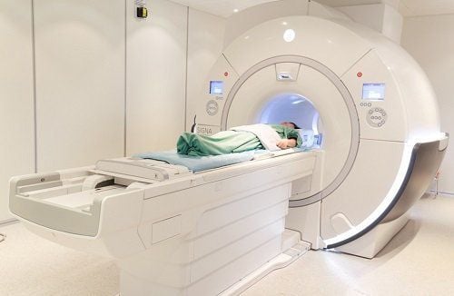
Cochlear Implants Clips in aneurysms Brain circuit Metal coils placed in the blood vessels First-generation defibrillators and pacemakers Once prepared, the patient will be placed on a dedicated imaging table that can be moved into the body of the device . The technician may use additional straps and splints to help the patient lie still and maintain his or her position during the scan. Devices containing coils capable of sending and receiving radio waves are moved to the pelvis, the area to be imaged, performing multiple runs (sequences) to obtain the images. Each sequence can last several minutes.
If the MRI uses contrast, the doctor or technician will insert an intravenous catheter into a vein in your arm to inject the necessary amount of contrast material into your body. At this time, the vascular system will appear more clearly.
Depending on the procedure and equipment used, the entire MRI scan is usually completed in 30 to 50 minutes and is painless. However, some patients find it uncomfortable to lie still for a long time. Others may feel claustrophobic while inside the MRI scanner. However, technicians can still see, hear and talk to the patient at all times using the two-way intercom. Therefore, if feeling too uncomfortable, the patient can give the signal to pause the MRI scan.
After completing the pelvic MRI, the results will be analyzed by experienced radiologists. Combined with the data from the physical examination and the results of other tests, the disease in the pelvis will be identified and a treatment plan will be devised.
In summary, MRI scans, using magnets and radio waves, are a safe, non-invasive means of obtaining images of the inside of the body without the need for surgical incisions. Accordingly, pelvic MRI is a useful survey tool to help doctors evaluate diseases of organs, blood vessels and other tissues in the pelvis. From there, common conditions in this area such as unexplained abdominal pain, the spread of some cancers... will be detected and treated early.

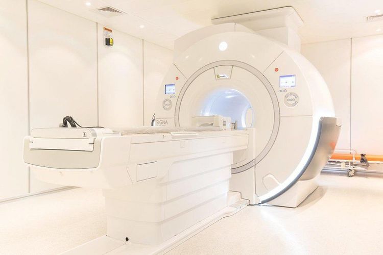
Hệ thống máy chụp cộng hưởng từ MRI 3.0 Tesla hiện đại tại Bệnh viện Vinmec Hải Phòng
High imaging technology, first-class safety by accuracy, non-invasiveness and no radiation High image quality, allowing doctors to evaluate comprehensive, does not miss even the smallest lesions in organs Minimize noise, create the most comfort for the patient when taking pictures, reduce stress, this helps to improve image quality and shorten Maximum scanning time MRI with Silent technology is especially suitable for the elderly and children, people with weak health, patients undergoing surgery... MRI at Vinmec can take vascular reconstruction. 3-D blood without injecting magnetic contrast, can take reconstruction and handle motion disturbances of the patient Maximum support for specialists to examine in-depth, detect diseases of the skull and neck region , spine, musculoskeletal, cardiovascular and oncology most precisely.
Please dial HOTLINE for more information or register for an appointment HERE. Download MyVinmec app to make appointments faster and to manage your bookings easily.


