This is an automatically translated article.
The article was written by Doctor of Emergency Department - Vinmec Phu Quoc International General HospitalUrine test is one of the very important health check and evaluation activities, which is indicated in general health examination and screening for many other diseases. Each parameter in the urine test has a certain meaning to reflect the health status of the body.
2. Changes in the properties of urine
2.1. Change of physical properties 2.1. 1. 24-hour urine volume The 24-hour urine volume depends on many factors, such as diet, fluid intake, ambient temperature, climatic conditions, physical activity, and use of diuretics. The average urine output in a normal person is 800-1800ml/24 hours.
+ Polyuria: when urine output is >2000ml/24 hours
+ Oliguria (little urination): when urine output is from 100 to less than 500ml/24 hours
+Anuria: when urine output is <100ml/24 hours
Physiological polyuria can be encountered when drinking too much water, infusing a lot of fluids, taking diuretics. But polyuria is commonly associated with pathologic states, such as diabetes mellitus, diabetes insipidus, recurrent episodes of acute renal failure, chronic tubular disease, the first days after transplantation, and the convalescent phase. recovery from some infectious diseases.
Physiological oliguria can be encountered due to drinking less water, sweating a lot, working in hot and humid climates without having enough water. Pathological oliguria may be caused by high fever, vomiting, diarrhea, hypotension, severe heart failure, acute or chronic renal failure, acute or chronic glomerulonephritis.
Anuria, seen in acute renal failure, severe progression of chronic renal failure, end-stage renal failure. There is only pathological anuria without physiological anuria.

Thể tích nước tiểu trong 24 giờ phụ thuộc vào nhiều yếu tố
2.1.2. Urine color Urine is normally clear, colorless, or pale yellow to yellow, depending on how concentrated the urine is. Urine contains abnormal solutes that can be identified by a change in the color of the urine. The more dilute the urine, the lighter the color, the more concentrated the darker the color. Dilute urine is usually colorless, and concentrated urine is usually dark amber. It is important to correctly recognize the color and watch for the color change that occurs with prolonged urine storage. Recently passed urine (fresh urine) may be clear, cloudy, cloudy, or pink in color.
+ Cloudy urine can be caused by:
Purulent urine: purulent urine, when left for a long time, settles into 3 layers, the top layer is clear, the middle layer is cloudy with pus lines, the lower layer is cells. Laboratory tests show many white blood cells, pus cells are degenerated polymorphonuclear leukocytes, possibly including bacteria. Urine has a lot of phosphate deposits (when the urine is alkaline), let the phosphate residue settle to the bottom, heat the insoluble residue, but add acid to the base to dissolve, the test shows many phosphate crystals. Urate has many amorphous urate residues (when the urine is acidic): let the urate residue settle to the bottom, heat or add alkaline solution to the residue to dissolve, the test shows many urate residues. Urine has chylothorax: if the amount of chylo is small, the urine will be cloudy like tapioca starch water, so it won't settle for a long time. If the amount of chlorophyll is too long, it will solidify like jelly, when the ether is added to the urine, it becomes clear. If there is blood in it, it is called hematuria, the urine is cloudy and red like meat washing water. + Urine is light red to dark brown: gross hematuria, the red blood cells will settle to the bottom for a long time, the test shows many red blood cells in the urine.
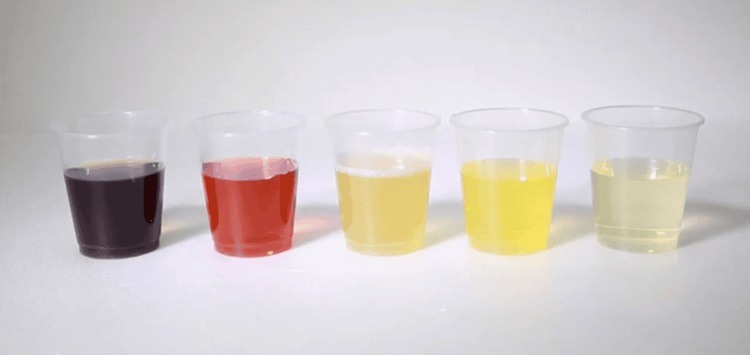
Màu sắc nước tiểu phản ánh một phần tình trạng sức khỏe của cơ thể
+ Urine is red-brown to brown: hemoglobinuria or myoglobinuria. For a long time, the urine becomes darker and does not settle. Tests for hemoglobin or myoglobin.
+ Other abnormal colors of urine: Red-yellow urine due to porphyrins, dark yellow due to bile or drugs (after taking tetracyclines), blue due to taking drugs such as mictasol blue.
Table 1: Some abnormal colors of urine and causes
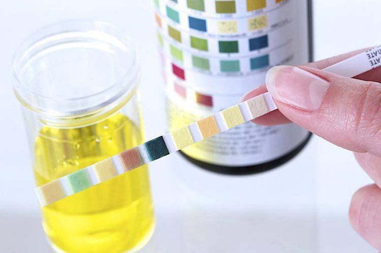
2.1.3. Urine pH Normal fresh urine can be neutral, acidic or alkaline. Urine pH can vary from 4.6 - 8, with an average of 5.5 - 6.5. Urine that has been kept for a long time has an alkaline reaction because urea is broken down and ammonia is released.
Urine pH test should be done soon after sampling, if a person's fresh urine has a frequent acid or alkaline reaction, it may be due to one of the following disorders:
+ Urine has a persistent acid reaction. :
Renal tuberculosis, Prolonged fever. Metabolic acidosis due to severe diarrhea, starvation, diabetic ketoacidosis, uremia and some cases of toxicity. Respiratory acidosis due to hypoventilation Methanol toxicity due to formic acid formation Metabolic disorders: phenil ketoneuria, ancaptonuria + Urine with prolonged alkaline reaction:
Genitourinary and urinary infections Metabolic alkalosis Use too much bicarbonate or other alkaloids Respiratory alkalosis due to hyperventilation Taking carbonic anhydrase inhibitor diuretics (diamox)
pH nước tiểu có thể thay đổi từ 4,6 - 8, trung bình là 5,5 - 6,5
2.1.4. Urine density and urine osmolality In a normal person, the body has a great adaptability to regulate water and electrolyte balance, so urine density and osmolality fluctuate within a very wide range. When drinking a lot of water, giving a lot of fluids, the kidneys increase the excretion of free water, making the urine dilute, so the density and urine osmolality are low, the density can be down to 1.003 or the osmolality 50 mOsm/kg H2O. When fasting, dehydration, the kidneys increase the reabsorption of free water, making the urine concentrated, the urine density can be as high as 1.030 or the osmolality 1200 mOsm/kg H2O.
In a normal person, drinking an average amount of water (according to demand) 24-hour urine density fluctuates on average from 1.015-1.025, osmolality ranges from 400-800 mOsm/kg. Urine samples during the day have a very large change in density or osmolality, depending on the time of eating and physical activity. Night urine samples usually have a higher density or osmolality than daytime urine samples, with early morning urine samples having the highest density. Urine specific gravity is usually high when there are high molecular weight solutes in the urine, such as glucose, contrast agents, magnesium, sulphate, or mannitol.
+ Decreased urine density and osmolality seen in the following diseases:
Tubulointerstitial diseases: acute pyelonephritis or chronic pyelonephritis, chronic interstitial nephritis, polycystic kidney disease return of acute renal failure, or the first days after kidney transplantation. In chronic renal failure due to chronic glomerulonephritis, urine density and osmolality are decreased, urine samples of the same day have low densities, range from 1.008 to 1.013, or osmolality ranges from 250 to 350 mOsm/kg H2O, regardless of the amount of water in the body. In chronic renal failure due to tubulointerstitial disease, urine osmolality and density decline early before renal failure, and in the presence of renal failure urine samples also have low copper density. With diabetes insipidus, urine attenuation and osmolality were very low (≤ 1,004, or <100 mOsm/kg).

Uống nhiều nước làm cho tỉ trọng và độ thẩm thấu nước tiểu thấp
Urine density and osmolality reflect the kidney's ability to concentrate urine. The density and method of measuring urine density have many influencing factors, which can easily falsify the results, making an inaccurate assessment of the kidney's ability to concentrate urine, for example, when the urine has a lot of protein or glucose. urine volume increased significantly.
Urine osmolality appears to have more advantages than urine density in assessing the ability of the kidneys to concentrate urine. Unlike urine density, urine osmolality does not depend on the molecular weight of the urinary solutes, but only on the number of "particle" solutes present in the urine. That more closely reflects the kidney's ability to concentrate urine than it does urine density. Urine osmolality measurement also allows the calculation of a number of parameters, such as osmotic clearance, free water clearance, urine osmolality and blood osmolality, which are very important parameters. Valuable in assessing the ability of the kidney to concentrate urine. Therefore, the osmolality method has gradually been used to replace the urine density method in assessing the ability of the kidneys to concentrate urine. If urine has no glucose or protein, urine osmolality is strongly correlated with urine density (r = 0.97, Ha Hoang Kiem 1994). Urine osmolality ranges from 40 to 1200 mOsm/kg. Nocturnal urine samples are usually more osmolal than daytime urine samples. If a urine sample of 75c at any time of day, or an early morning urine sample, has an osmolality of 600 mOsm/kg H2O, the kidney's ability to concentrate urine can be assumed to be normal. (Ha Hoang Kiem 1998).
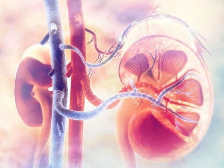
2.2. Change of biochemical components of urine 2.2.1. Proteinuria In the normal person, only a very small amount of protein in the plasma is filtered by the glomeruli, but is completely or almost completely reabsorbed by the proximal tubule cells. Only very little protein is excreted in the urine in a day, which is not detected by conventional biochemical tests and is negative. Currently, with radioimmunoassay (RIA) technology, it is found that in normal people, about <30 mg of protein is excreted in the urine in a day, of which albumin 9 mg, IgG 3 mg, and natural light chains. due to 3.3 mg. If the urine has ≥30mg of protein/24 hours, it is an indicator of kidney damage (before testing, it is necessary to make sure that the urine is not mixed with blood or pus, because these substances contain proteins, the water must be filtered. pee before the test)
Protein trong huyết tương được hấp thu gần hoàn toàn qua cầu thận
+ If the amount of protein in the urine is from 30 to 299 mg/24 hours, it is called microalbuminuria. With this amount of proteinuria, conventional biochemical methods cannot detect it, but must be tested by radioimmunoassay, now there is a rapid test strip for microalbuminuria.
Microalbuminuria is a parameter used to detect kidney damage early, such as in diabetes, high blood pressure...
+ When urine protein is ≥300 mg/24 hours, biological tests routine test results are positive. When there is frequent proteinuria, it is definitely pathological proteinuria.
+ Some cases have protein in the urine, but no kidney damage, it is necessary to distinguish:
Proteinuria due to standing position: can be seen at age < 20, if there is proteinuria age > 30 is usually not postural proteinuria. To differentiate, let the patient rest for at least 8 hours (overnight), re-test will show negative proteinuria, let the patient stand for more than 1 hour, proteinuria is positive again, but lying down is negative. Protein from standing posture is not associated with erythrocytosis, or other symptoms of kidney disease. However, individuals with orthostatic proteinuria need close and long-term monitoring. Some studies show that, after many years, some people with orthostatic proteinuria have developed kidney disease, so the concept of orthostatic proteinuria as benign proteinuria according to some authors needs to be reconsidered. Bence-Jones urine: also known as heat-soluble proteinuria, or paraprotein. Bence-Jones proteinuria will precipitate cloudy urine when heated to 40 C-60 C, but when heated to 100 C, Bence-Jones protein will dissolve to make urine clear again, but let cool to 40 C-60 C The urine is cloudy again due to precipitation of Bence Jones protein, the nature of Bence Jones protein is the kappa or lamda light chain of immunoglobulins produced by cancer cells, with a molecular weight of 60 Daltons, so it is easily filtered by the glomeruli. When the reabsorption threshold of the proximal tubule is exceeded, it is excreted in the urine. Bence-Jones proteins are seen in multiple myeloma (Kahler's disease), cancer, and some patients with Waldenstrom's disease (about 10% of patients). Proteinuria caused by a number of diseases other than kidney disease such as congestive heart failure in the presence of oliguria, septic fever, traumatic brain injury and meningeal bleeding. Proteinuria due to the above diseases appears only temporarily during the illness. Proteinuria in first-time pregnant women: first-time pregnant women have proteinuria in the third trimester of pregnancy, accompanied by hypertension and edema, which is a sign of pregnancy toxicity, if severe, pregnant women may have eclampsia, stillbirth. A few weeks after delivery, the symptoms will disappear and the proteinuria will be negative. If proteinuria persists after parturition, pre-existing kidney disease is likely. If proteinuria is present in the early months of pregnancy, it is also more likely to have pre-existing kidney disease. Women who miscarry in their first pregnancy can still have gestational toxicity in subsequent pregnancies.

Protein niệu ở người có thai lần đầu sẽ tự hết sau khi đẻ vài tuần
+ Proteinuria due to kidney disease:
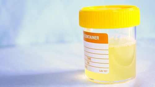
When there is frequent proteinuria, it is certain that pathological proteinuria is usually a kidney disease. The amount of proteinuria/24 hours has pathological value whether in the glomerulus or in the renal tubule. Low proteinuria (<1g 24 hours): Low proteinuria often indicates pathology of the renal tubules such as acute pyelonephritis or chronic interstitial nephritis, renal vascular fibrosis (due to hypertension), polyuria follicle. Proteinuria due to tubulointerstitial disease usually has the same ratio of albumin to globulin as in serum (A/G = 1.2-1.5). High amount of proteinuria: usually a manifestation of glomerular disease, chronic glomerulonephritis without nephrotic syndrome, the amount of proteinuria is usually in the range of 2-3g/24 hours. If nephrotic syndrome is present, proteinuria is high (3-5g/24 hours), blood protein is decreased (<60g/l), blood lipids are increased, edema, and lipids are present in the urine. The proteinuria component in nephrotic syndrome due to minimal glomerulonephritis or membranous glomerulonephritis, is mostly albumin (albumin accounts for 80% of proteinuria), globulin accounts for only 20%, of which β globulin is the most abundant. This is called selective proteinuria (the urine globulin/albumin ratio is called the selectivity index, normal ≥1.0. If the ratio is above 1.0, it is selective proteinuria, if < 0). ,5 is highly selective, and the lower the number, the higher the selectivity). 2.2.2. Hemoglobinuria Hemoglobinuria is also known as hematuria. Normally, in plasma, there is only 1-4 mg of hemoglobin in 100 ml of serum, in urine there is no hemoglobin. When blood hemoglobin levels increase, hemoglobinuria is caused. When there is acute hemolysis due to the wrong blood type, venomous snakebite that causes hemolysis, hemoglobinuria from malignant malaria, hemoglobin from the red blood cells is released into the plasma in large quantities, the liver does not convert promptly converted to conjugated bilirubin (indirect bilirubin), to a certain extent is excreted unchanged in the urine. Hemoglobinuria can be seen in the following diseases:
+ Malaria hematuria, often caused by Plasmodium Falciparum
+ Bacterial infections, usually caused by Clostridium perfringens
+ Chemical poisoning such as arsenic, snake venom, some drugs like amphotericin
Hemoglobin trong huyết tương quá nhiều sẽ được bài xuất nguyên dạng ra nước tiểu
+ Transfusion of the wrong blood group causes acute hemolysis
+ Paroxysmal hemoglobinuria due to cold (rare): after cold infection, the patient has chills, abdominal pain, low back pain, muscle pain and hemoglobinuria.
+ Paroxysmal nocturnal hemoglobinuria, seen in patients with Marchiafara-Micheli syndrome, consists of episodes of paroxysmal nocturnal hemoglobinuria, usually at age 30-40.
+ Diarrhea in hemoglobin due to abnormal and prolonged exercise, such as long distance running in people who are not used to exercise. After exercise, hemoglobin in urine, red blood cells in the blood have a normal shape.
+ Drug-induced hemoglobinuria, seen in G6PD deficiency.
2.2.3. Myoglobinuria Myoglobin is a breakdown product of muscle, and myoglobinuria occurs when a lot of muscle is crushed, as in burial syndrome. Congenital primary myoglobinuria can sometimes be familial. Manifestations of muscle pain, low proteinuria, erythrocytosis, myoglobinuria, possibly anuria.
2.2.4. Chrysanthemum urine Normal urine does not have chylous, if there is chylous, the urine is milky white, or like tapioca starch juice. If the amount of chlorophyll is a lot of urine left in the test tube for a long time, it will solidify like a clear white jelly. It is also possible to have both chylous urine and blood in the urine, and the urine is milk chocolate color. Chyluria occurs when there is a fistula from the lymphatic system into the renal or urinary system, often the fistula enters the calyx or renal pelvis. Lymphatic-urinary fistulas can be detected by radiography of the lymphatic system, or retrograde pyelonephritis. Chilli is mainly composed of triglycerides, cholesterol, and protein. Because it contains a lot of lipids, the urine is milky white. The higher the fat in the diet, the more severe the chylous diarrhea.
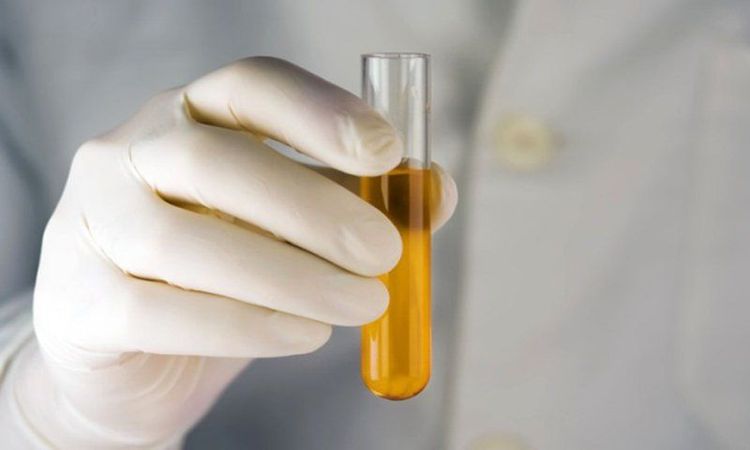
2.2.5. Lipiduria In a normal person, about 12 mg of lipids per day are excreted in the urine, including 3.1% fatty acids, 0.25% phospholipids, 0.21% triglycerides, and 0.05% cholesterol. Almost no cholesterol.
Mỗi ngày có khoảng 12 mg lipid được đào thải qua nước tiểu
In nephrotic syndrome, the increase in lipids in the urine can be more than 0.5g/24 hours, in which free fatty acids account for the highest proportion, phospholipids and cholesterol in the urine are increased and the urinary triglycerides are not increased.
Cholesterol esters in the urine of patients with schizophrenia form globular, cross-shaped bodies that glisten under polarized black-field microscopy light, called photorefractive bodies. Urinary optical birefringence is a valuable parameter in the diagnosis of nephrotic syndrome.
Increased lipiduria is also seen in the end of normal pregnancy, in patients with diabetes, biliary cirrhosis, hypothyroidism, end-stage renal failure, but not as high as nephrotic syndrome and can not be birefringent. optical.
2.2.6. Porphyrinuria Porphyrinuria is seen in acute porphyria, which is the manifestation of a familial disease of metabolic disorder that causes excessive porphyrin production. Depending on where porphyrin is produced, people are divided into three groups:
+ Liver porphyria
+ Porphyrin caused by red blood cell formation.
+ Combined porphyria
When one of the enzymes involved in heme synthesis is lacking, one of the above groups of porphyrinuria will occur. The disease often comes on suddenly, after an infection, or after taking certain medications. Clinical manifestations include acute pain, motor paralysis sometimes even respiratory paralysis, appearing after several days of abdominal pain. The urine is sometimes dark red in color after several hours of urinating, and the urinalysis shows porphyria, which is a medical emergency.
If you have a need for consultation and examination at Vinmec Hospitals under the nationwide health system, please book an appointment on the website for service.
Please dial HOTLINE for more information or register for an appointment HERE. Download MyVinmec app to make appointments faster and to manage your bookings easily.


