This is an automatically translated article.
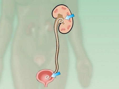
JJ Stent (Double-J) is a hollow plastic tube 24-30cm long, specially designed for insertion into the ureter. Placement of stents under enhanced radiographs is an effective method to treat ureteral strictures, helping to reestablish urinary tract excretory flow.
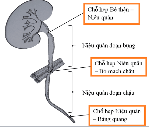
1. Overview of ureteral stricture
The ureter is the natural tube that carries urine from the kidney to the bladder. Ureteral stricture is one of the common pathologies in the pathology of the urinary system, which can cause hydronephrosis upstream and lead to renal failure. Endoscopic retrograde ureteral stenting (Double-J) is often the preferred choice because of its minimal invasiveness.Effect of ureteral stricture treatment with Stent:
Insertion of a ureteral stent (Double-J) helps urine to flow from the kidney down to the bladder. Helps the kidneys continue to work and not be damaged by the blockage. Reduce the risk of infection, limit severe renal colic pain when the kidneys are not drained well. Protect the ureter, help the injured ureter gradually heal, prevent the risk of ureter narrowing when the wound heals.

Đặt stent niệu quản (Double-J) ngăn ngừa những cơn đau quặn thận
2. Causes of Congestion
Place a ureteral stent (Double-J) when there is a blockage in the ureter or kidney, helping to improve the obstruction. Common causes of obstruction are:
Ureteral stones : Stones form in the kidney, then impacted, move down the ureter and cause obstruction. Depending on the size of the stone, small stones can pass out on their own through the urinary tract, but some remain stuck, causing obstruction. Tumors: Both benign and malignant tumors can cause ureteral obstruction, requiring stenting to clear the blockage in the kidney. Stenosis: Narrowing can be caused by scarring that causes narrowing and obstruction of the ureter. Injury to the ureter: Injuries that occur during ureteroscopy (eg, endoscopic bowel surgery, treatment of stones or performing biopsies, obstetrics and gynecological procedures..) or due to direct injury to the ureter. due to penetrating wound.
3. Indications and contraindications for ureteral stenting under enhanced radiography
Indications:
Urethral stricture due to many different causes. Ureteral fistula Before percutaneous lithotripsy (>15mm large lithotripsy or solitary kidney) After ureteroplasty and retrograde ureteroscopy. Contraindications:
Bladder diverticulum Small bladder syndrome Urinary incontinence Acute urinary tract infection Hemorrhage of the urinary tract following percutaneous pyelonephritis
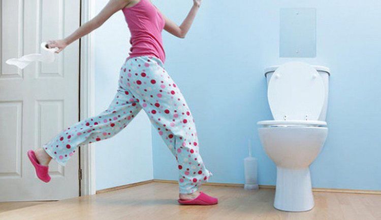
Bệnh nhân tiểu không tự chủ chống chỉ định thực hiện
4. Procedure for placing ureteral stents under X-ray to treat ureteral stenosis
4.1 Preparation
To perform the procedure of ureteral stenting (Double-J) under enhanced radiography, it is necessary to prepare:
Implementation team:
Specialist doctor, ancillary doctor Radiology technician Nursing Doctor/technician General anesthesia technician Means of use:
Fluoroscopy. Film, film printers and image storage systems. Lead vest and apron for protection from X-rays. Ultrasonic flat and curved transducer. Sterile plastic bag wrapped with ultrasound transducer Medicine: Includes local anesthetic, general anesthetic (if necessary), water-soluble iodine contrast agent, antiseptic solution for skin and mucous membranes.
Common medical supplies: 5.10ml syringe, distilled water (physiological saline), surgical clothing; sterile intervention kit (knife, scissors, forceps, instrument tray, etc.), cotton gauze, surgical tape; medicine box and accident first aid box.
Special medical supplies:
Kim Chiba and corresponding leads Standard angiography catheter 4-5F Standard lead 0.035'' Pigtail drain. Ureteral catheter (Double J) 6-8F length 22-28cm. Drug pump connection Suture to fix the catheter. Patients need to prepare:
Be explained in detail about the procedure to coordinate with the doctor. Perform clinical examination before the procedure. Fasting, drinking before 6 hours. Do not drink more than 50ml of water. In the intervention room: the patient is assigned to lie on his or her side, depending on the location of the drainage. The doctor installs a machine to monitor breathing, pulse, electrocardiogram, blood pressure, and oxygen saturation in the peripheral blood. Approximately 2-7 days prior to percutaneous ureteral (Double-J) stenting, a percutaneous pyelonephritis should be placed in place to resolve obstructive pyelonephritis and minimize the risk of renal failure. urinary tract bleeding.

Bệnh nhân cần nhịn ăn, uống trước 6 tiếng
4.2 Ureteral stenting procedure (Double-J)
Steps to treat ureteral stenosis under enhanced X-ray:
Step 1: Take pyelogram - ureter downstream
Through the catheter to drain the pyelonephritis through the skin, inject contrast to take pictures of the pyel, ureter . Assess the extent and location of the blockage. Step 2: Create an entrance to the urinary tract
Insert the 0.035'' lead into the renal pelvis and ureter to withdraw the drainage catheter (Pigtail). Insert the sheath 6-8Fr into the renal pelvis according to the lead. Step 3: Approach the injured area
Use a catheter and wire to go from the renal pelvis through the ureter down to the bladder. Replace the standard lead with a stiff wire (Stiff wire), remove the catheter. Step 4: Insert the ureteral stent (Double J)
Insert the ureteral stent into the renal pelvis, ureter and bladder by rigid lead. Then withdraw the lead back to the renal pelvis. Place a catheter to drain the renal pelvis through the skin. Inject the contrast agent into the renal pelvis, locate the superior end of the ureteral stent, and assess the stent's circulation. Step 5: Place percutaneous pyelonephritis
Fix the position and lock the percutaneous pyelonephritis catheter. After 24-48 hours, assess that the ureteral stent is working well and is not obstructed, then remove the percutaneous pyelonephritis catheter. Step 6: Evaluation of the results
Regarding the position, the JJ ureteral stent needs to have the lower end bent in the bladder and the upper end bent in the renal pelvis. In terms of function, when pumping drugs from the renal pelvis to the bladder, there must be signs of circulation. Do not drain the contrast agent out of the urinary tract into the abdomen or into the retroperitoneal space.
5. Attention when placing ureteral stents (Double-J)
The patient can still do normal activities (work, play sports, travel, have sex...) but will feel tired and uncomfortable more quickly. In addition, the patient may have a need to urinate more than usual.
While having a ureteral stent (Double-J), the patient should drink at least 1.5-2 liters of water per day. Monitor and notify your doctor immediately if you notice unpleasant side effects such as:
Frequent and intolerable lower back pain due to stents. Signs of a urinary tract infection (fever, cold, malaise, burning pain when urinating). Stent falls out. The frequency of hematuria increased significantly.

Sau đặt stent niệu quản (Double-J) , người bệnh cần uống nhiều nước
6. Complications after the procedure and treatment direction
After the procedure, many patients can tolerate stents easily. Others have some side effects such as:
Infection:
Foot drainage infection: cleaning, dressing change and local antiseptic. Urinary tract infections: systemic antibiotics are indicated. Sepsis: conduct specialist consultation. Urinary frequency: A ureteral stent (Double-J) can irritate the bladder and cause a person to urinate more often. These symptoms are completely normal and should improve after taking the medicine.
Stent deviation out of position: In this case, the Stent usually moves down to the bladder and causes more severe symptoms such as pain, discomfort in the hip, back, bladder, groin, penis... with urinary frequency and hematuria.
Bleeding in the renal pelvis, ureter, retroperitoneum:
Assess by the amount of urine excreted through the drain and drain. Use your hands to press at the lumbar hole with a drain for 15-20 minutes. If bleeding persists, move to intervention room to replace with larger size drain. After replacing the drain tube, if bleeding is still done, perform vascular intervention. These side effects may persist only for the first few days or weeks after ureteral stenting (Double-J). Some patients find that these symptoms persist for as long as the stent is in their system. These side effects usually go away completely after the stent is removed.
The ureteral stent (Double-J) is removed using a cystoscope after local or general anesthesia. A special flexible, telescope-shaped endoscope is inserted into the urethra, and the stent is picked up and withdrawn. Stents are usually not kept for more than 3 months in the body.
Vinmec International General Hospital with a system of modern facilities, medical equipment and a team of experts and doctors with many years of experience in medical examination and treatment, patients can rest assured to visit. examination and treatment at the Hospital.
To register for examination and treatment at Vinmec International General Hospital, you can contact Vinmec Health System nationwide, or register online HERE.


