This is an automatically translated article.
The article was professionally consulted with Master, Doctor Le Anh Viet - Radiologist - Department of Diagnostic Imaging and Nuclear Medicine - Vinmec Times City International Hospital.Cirrhosis is one of the common liver damage caused by hepatitis. Currently, ultrasound measuring liver fibrosis is a necessary diagnosis, in which, ARFI ultrasound technique is a modern ultrasound technique, allowing to assess the degree of cirrhosis with high accuracy. .
1. Overview of cirrhosis of the liver
1.1 What is cirrhosis of the liver?
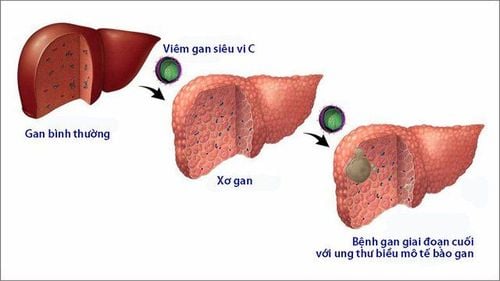
Cirrhosis is a long-term damage to liver cells, scar tissue in the liver is continuously formed to replace damaged liver tissue cells. The more scar tissue the liver has, the more it will impede blood circulation to the liver, thereby severely reducing liver function.1.2 What are the stages of cirrhosis of the liver?
Cirrhosis has 4 stages:Cirrhosis in the early stages - Cirrhosis F1 : Liver cells and tissues begin to be damaged. At this stage, the disease has no typical clinical symptoms, the patient may feel mild abdominal pain, loss of appetite, fatigue, low fever, ... At this time, liver function has not deteriorated, so if possible If the liver fibrosis is measured by ultrasound in time, the recovery rate is high. Cirrhosis stage 2 - F2 cirrhosis (also known as liver fibrosis): Liver cells and tissues are more damaged and begin to form scar tissue and tissue fibrosis. At this stage, the patient has symptoms similar to the first stage, but with a higher degree. In addition, it is also possible to detect symptoms of jaundice, yellow eyes, ... due to a disorder in the metabolism of bilirubin in the liver. At this time, liver function is significantly reduced. Cirrhosis stage 3 - Cirrhosis F3: The number of cirrhosis liver cells is increasing and causing serious liver dysfunction. Typical symptoms of the disease such as jaundice, yellow eyes, body fatigue, edema, ... appear clearly. End stage cirrhosis - F4 cirrhosis: Liver cells and tissues are completely damaged and liver function is lost. At this stage, the disease can cause dangerous complications such as cirrhosis, ascites and abdominal distension, or progress to liver cancer. Periodic health examination, including ultrasound to measure liver fibrosis is the best measure to help detect and prevent cirrhosis from progressing.
2. The role of ultrasound in measuring liver fibrosis

Evaluation of liver fibrosis helps prevent complications caused by advanced cirrhosis such as decompensated cirrhosis, liver cancer. If diagnosed, evaluated, and treated early, severe cirrhosis can be prevented with a high recovery rate after treatment. Specifically, the role of ultrasound to measure liver fibrosis is as follows:
Assess severity, cirrhosis status. Indication of treatment is appropriate to the extent and condition of the disease. Monitor progress and assess the patient's ability to respond to treatment. Provide early disease prognosis, help prevent cirrhosis from progressing.
3. Ultrasound technique to measure liver fibrosis ARFI
Acoustic radiation force impulse imaging (ARFI) is an ultrasound technique that creates acoustic radiation impulses. This is a new elastographic method of choice first in the assessment of liver fibrosis, allowing an accurate diagnosis of the stage of acquired cirrhosis.
3.1 Principle of ultrasound technique to measure liver fibrosis ARFI
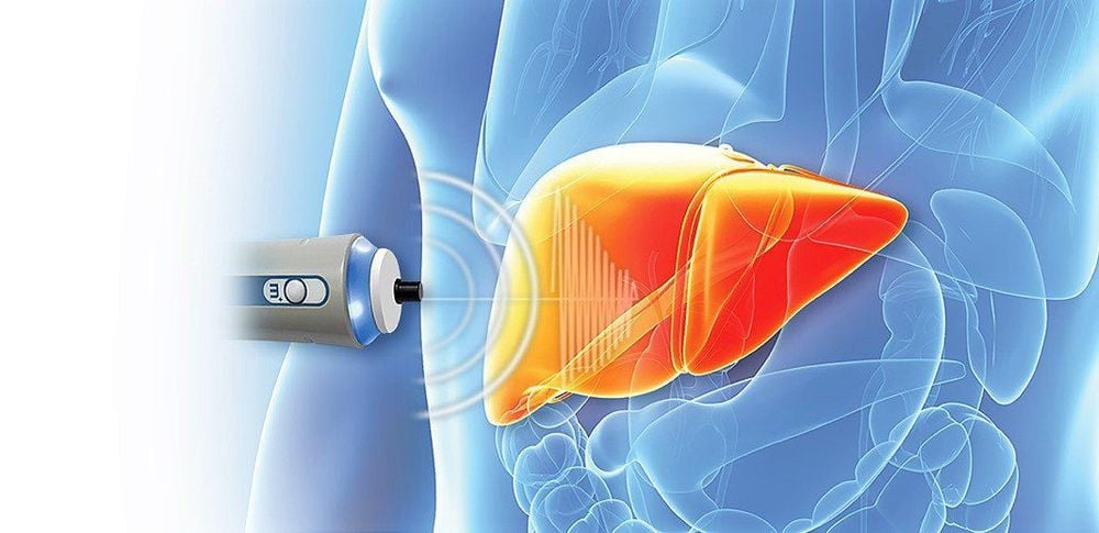
The principle of ultrasound technique to measure liver fibrosis ARFI is the operation of imaging by acoustic radiation pulses, allowing to examine the tissues and cells deep inside without having to press the transducer strongly.In a short time, using a push pulse, this technique mechanically stimulates the tissue in the selected ROI to generate shear waves perpendicular to the impulse. This technique measures the shear wave velocity using a conventional SA beam and tracks it on the same transducer.
3.2 Highlights of ARFI . ultrasound technique
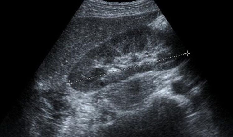
The ultrasound technique to measure liver fibrosis ARFI has the following outstanding features:Accurately assess the degree of liver fibrosis due to a set of standard parameters. The ARFI software is integrated and installed into the conventional ultrasound machine, allowing the simultaneous performance of standard liver ultrasound examination and measurement of liver fibrosis. Selected examination area, thereby avoiding blood vessels in the measurement area. Fibrometry can be performed in both lobes of the liver. The ultrasound image of the liver fibrosis recorded gives more information about the stiffness of the liver tumor, the tumor border - the tumor contour, helping to distinguish liver hemangiomas from malignant liver tumors. Allowing to be performed in many different patients such as those who are overweight, obese, narrow rib space or have a lot of fluid in the abdomen, Simple and easy operation, non-invasive and painless. The time to perform the ultrasound is fast and gives instant results. The ARFI technique can be used to measure liver stiffness at the same time as Doppler and B-mode ultrasound.
3.3 Value of ultrasound technique to measure liver fibrosis ARFI
The technique of ultrasound to measure liver fibrosis ARFI is an accurate technique and brings high value in the diagnosis and assessment of liver fibrosis (from significant fibrosis to severe fibrosis and cirrhosis). limitations of some other techniques such as being able to perform in obese patients, narrow flank space, ascites, with high sensitivity and specificity.The ARFI ultrasound technique is often combined with some other ultrasound techniques to increase the specificity in the diagnosis of liver fibrosis significantly.
In diagnostic imaging, ultrasound to measure liver fibrosis ARFI is an elastographic technique that is being used relatively much to evaluate non-invasive liver fibrosis in liver diseases such as:
Liver disease Chronic viral diseases (hepatitis A, B, C): Ultrasound provides valuable diagnostic results with high accuracy in the assessment of liver fibrosis from significant fibrosis, to severe fibrosis and cirrhosis. Non-alcoholic fatty liver disease: As with chronic viral liver disease, ultrasound measurement of liver fibrosis ARFI has high value in assessing the degree of liver fibrosis in nonalcoholic fatty liver disease. ARFI is a new elastographic ultrasound technique used in the assessment of liver fibrosis with high accuracy.

In particular, to improve service quality, Vinmec International General Hospital now has Hepatobiliary Screening packages, which help detect liver diseases at an early stage even when there are no symptoms. In addition, the comprehensive hepatobiliary screening package helps customers:
Assess the liver's ability to work through liver enzyme tests; Evaluation of bile function; vascular nutrition; Early screening for liver cancer; Perform tests such as Total blood cell analysis, blood clotting ability, screening for hepatitis B, C Assessment of hepatobiliary status through ultrasound images and diseases that have the potential to affect liver disease/cause liver disease. more severe liver disease In-depth analysis of parameters to evaluate hepatobiliary function through laboratory, subclinical; the risk of affecting the liver and early screening for hepatobiliary cancer. When the results of the examination are available, the patient will be consulted and treated intensively with a specialist in Gastroenterology.
Please dial HOTLINE for more information or register for an appointment HERE. Download MyVinmec app to make appointments faster and to manage your bookings easily.


