This is an automatically translated article.
The article was professionally consulted by Specialist Doctor II Khong Tien Dat, Doctor of Radiology - Department of Diagnostic Imaging - Vinmec Ha Long International Hospital and Specialist Doctor I Nguyen Truong Duc - Doctor Doctor of Radiology - Department of Diagnostic Imaging and Nuclear Medicine - Vinmec Times City International Hospital.1. What is Contrast Echocardiography?
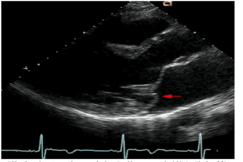
Contrast echocardiography is an ultrasound method combined with the injection of contrast material into the blood vessels to increase the ability to detect cardiac structures and flows in echocardiographic exploration (through the chest wall and through the esophagus).When conducting echocardiography, contrast material is injected into a vein and returned to the heart.
2. When is an ultrasound with intravenous contrast injection indicated?
When the doctor needs more information, assess the patient's health status:Patients with heart diseases such as: Congenital heart, need to find shunts between the heart chambers, blood vessels ...

3. Ultrasound procedure with intravenous contrast injection
To perform only need to use echocardiography with intravenous contrast, 1 nurse and a cardiologist to perform echocardiography.3.1 Prepare the media The facilities for the procedure include:
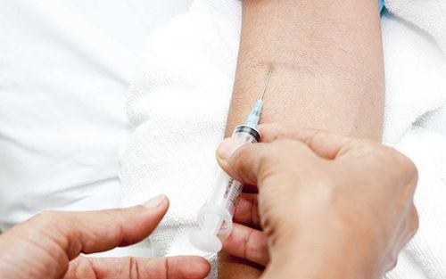
Ultrasound machine: Plain black and white with cardiac transducer. The heart probe of the ultrasound machine has a chip that can record video to analyze the image, the direction of the echo foam. Contrast Contrasts: Contrasts are solutions with properties that enhance the echocardiographic response to clarify cardiac structures, myocardial flow and perfusion, including self-made contrast agents and other substances. pre-manufactured muffler. Self-made contrast agents: Doctors will often use solutions for daily infusion to create acoustic foam, eg 0.9% NaCl solution, glucose, isotonic and hypertonic dextrose. 3.2 Steps to conduct echocardiography with intravenous contrast The nurse performs echocardiography as usual for the patient. A needle is then inserted into the vein in the arm (usually the left arm is chosen) and connected to the system.

Currently, Vinmec International General Hospital has also applied the ultrasound method with intravenous contrast injection in the indications of doctors in departments and clinics when further evaluation is required. Vinmec International General Hospital with international standards is a reliable destination to choose for examination and treatment.
Please dial HOTLINE for more information or register for an appointment HERE. Download MyVinmec app to make appointments faster and to manage your bookings easily.


