This is an automatically translated article.
The article was written by Specialist Doctor I Nguyen Dinh Hung - Radiologist, Department of Diagnostic Imaging - Vinmec Hai Phong International General Hospital.
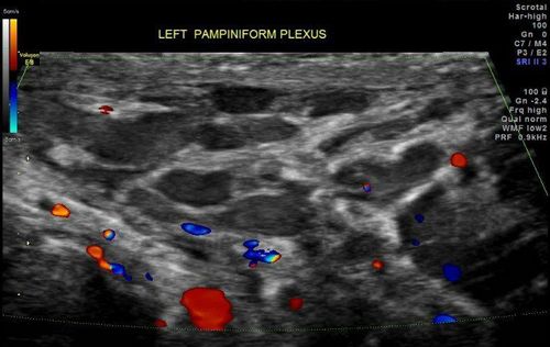
A varicocele is called a varicocele when at least one vein in the venous plexus is larger than 2.2 mm in diameter, with reflux causing the vein to enlarge after the patient stands up or performs the Valsalva maneuver.
1. What is varicocele?
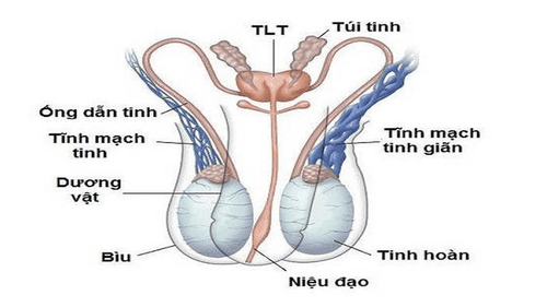
Varicocele is an abnormal dilation of the vas deferens and the plexus (normally, the varicocele of the vas deferens is about 2.2mm or less in diameter). Varicose veins are a very common and common disease in men. The main reason for patients to go to the doctor is pain in the groin area or infertility.
The disease rarely appears before puberty, in adolescence the incidence is from 8-19%, but since adolescence, according to the authors' general statistics, in about 15% of the male community .
However, in the male infertility group, the rate is much higher. Varicose veins are responsible for 15-25% of primary male infertility cases and 75-81% of secondary male infertility.

2. The seminal vein and the role of the seminal vein
The seminal vein originates from the testis and receives blood from the epididymis branches. The internal seminal vein is due to the veins joining together to form a tangled venous plexus, entangled in the spermatic cord, ascending and gathering into a large branch, the left empties into the renal vein and the right empties. into the abdominal vena cava. Veins have valves to prevent blood from backing up into the testicles.
Due to anatomical features, 90% of cases of varicocele are only found on one side, of which, up to 90% are found on the left side.
3. Ultrasound in the diagnosis of varicocele
Most cases of varicocele can be detected on physical examination when varicose veins are visible or palpable. In some cases, imaging aids are available, including ultrasound and often Doppler ultrasound.

4. Purpose of ultrasound in diagnosing varicocele
Color Doppler ultrasound of the testicular pedicle to accurately assess the extent of varicocele. Normally, the diameter of the seminiferous vein is less than 2 mm. Evaluation of abnormalities and associated lesions: Thrombosis, hydrocele, epididymal cyst. Doppler ultrasound: Evaluation of the reflux flow of the testicular veins. Detection of anatomical abnormalities of the left renal vein. Varicose veins classification.
Grade chart of varicocele measured on ultrasound before and after Valsalva maneuver
With a team of doctors with clinical and diagnostic experience, along with the new generation ultrasound system of GE: S8, E9 is equipped with the latest generation of Linear matrix transducers.
Vinmec International General Hospital is one of the hospitals that not only ensures professional quality with a team of leading medical doctors, modern equipment and technology, but also stands out for its examination and consultation services. comprehensive and professional medical consultation and treatment; civilized, polite, safe and sterile medical examination and treatment space. Customers when choosing to perform tests here can be completely assured of the accuracy of test results.
If you notice any unusual health problems, you should visit and consult with a specialist.
Please dial HOTLINE for more information or register for an appointment HERE. Download MyVinmec app to make appointments faster and to manage your bookings easily.


