This is an automatically translated article.
The article was professionally consulted by Doctor General Internal Medicine Doctor - Department of Medical Examination & Internal Medicine - Vinmec Hai Phong International General Hospital.
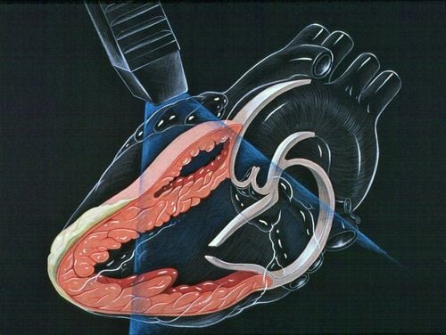
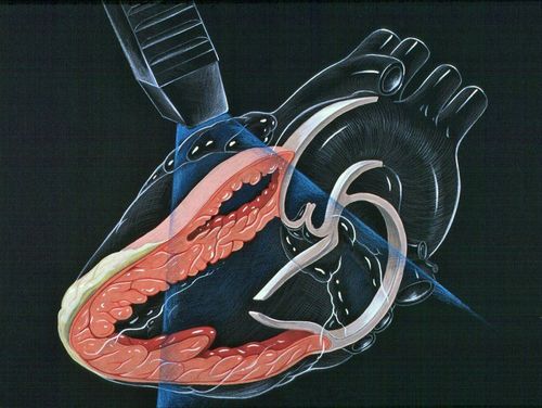
Doppler echocardiography is one of the imaging techniques used in cardiology, the main purpose is to observe the movement of the heart, the function of the heart muscle and the heart valves, diagnose and monitor the heart. effective in cardiovascular diseases. The Doppler echocardiography procedure is usually quite quick and can be done in a dedicated ultrasound room or emergency ultrasound at the bedside.
1. What is doppler echocardiography?
Doppler echocardiography is a noninvasive method that helps to probe the heart and great vessels in the mediastinum with a probe placed outside the patient's chest wall. The principle of this technique is to use high-frequency ultrasound waves (frequency 3-5 MHz) to probe, to observe and obtain images of the heart, movements or related structures. heart. Based on the images obtained, doctors can make an initial assessment of abnormalities of the heart muscle or of the heart valves.
2. What are the parameters on the echocardiogram obtained?
During an echocardiogram, your doctor will use transducers that move around your chest or esophagus. The ultrasound process will help the doctor easily observe and evaluate the parameters showing the function of the heart such as:
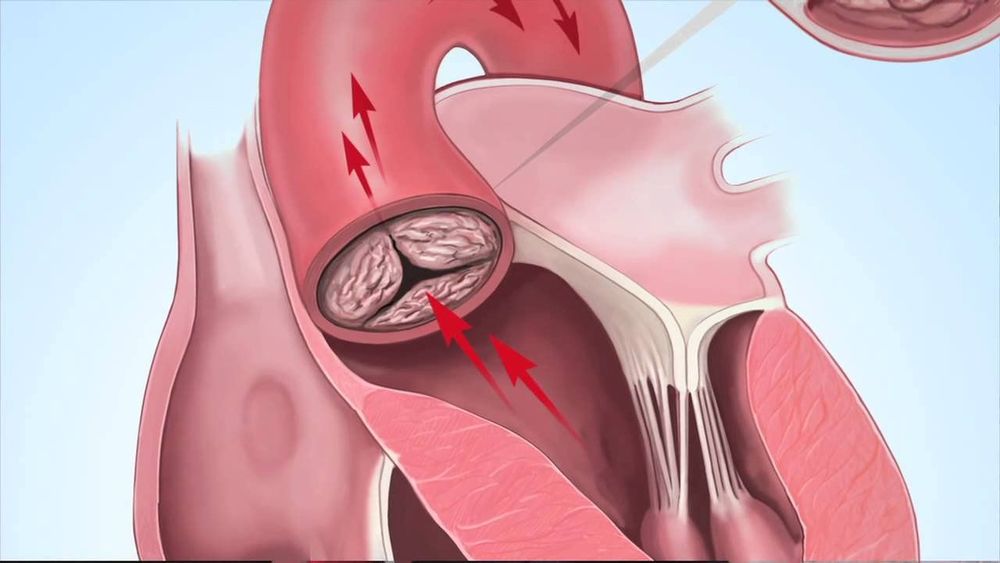
Shape, size, activity status, contraction of the heart. Size and pumping of the valves and walls of the heart. Check the heartbeat. The pumping power of the heart and the volume of blood pumped to other organs of the body, the pumping power per minute and the average value. Detect abnormal signs or detect damage to the heart such as: open heart valve, wide or narrow heart valve, abnormal blood clot in the heart, ....
3. Diseases that require echocardiography
Hypertension Ischemic cardiomyopathy or evaluation of cases of chest pain Heart rhythm disorders Heart valve disease Pericardial disease Evaluating cases of dyspnea Chest aortic disease Congenital heart diseases Follow up in heart surgery, resuscitation. Health check-ups and screenings in the community.
4. Doppler echocardiography procedure
Prior to Ultrasound When a Doppler echocardiogram is indicated, there is essentially no special preparation for the patient and no pre-ultrasound screening tests are required. Patients can eat and drink normally before the procedure.
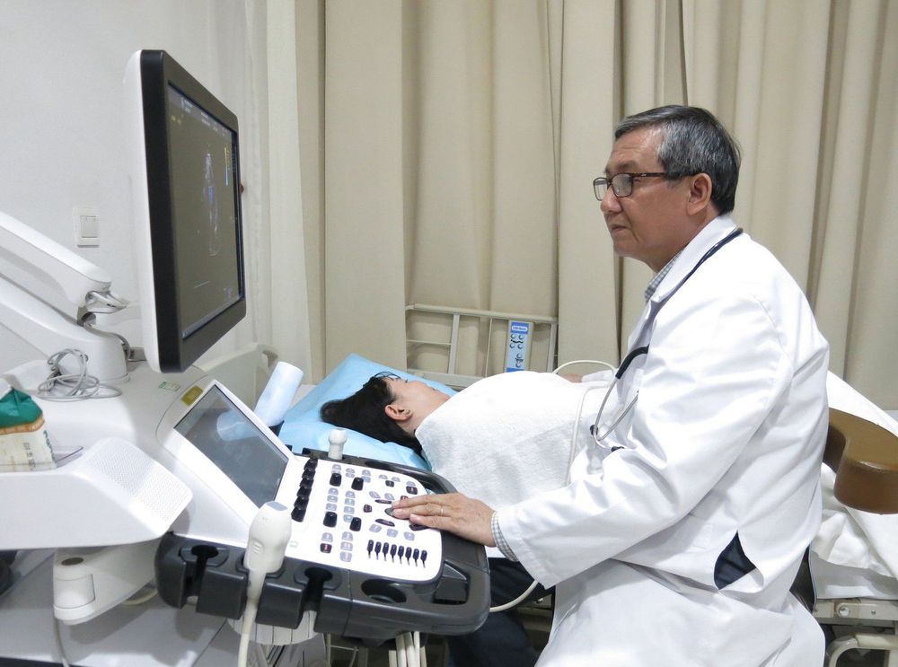
Patient position: Lie on your back, slightly leaning to the left, lying in a resting state. The patient had an electrocardiogram concurrently during Doppler echocardiography. Performer: Sitting on the right side of the sonographer, the right hand holds the transducer, the left hand adjusts the buttons of the machine.
During echocardiography The transducer is placed at the left sternal border, in the third or fourth intercostal space. The transducer will create an angle of 80 - 90 degrees with the patient's thoracic plane. Ultrasound waves are projected perpendicular to the heart structure, helping to measure the thickness and width of these structures.
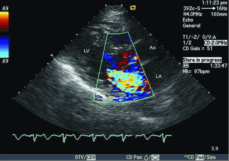
Cardiologist or radiologist will directly perform doppler echocardiography for the patient. The doctor can adjust the transducer to get different viewing angles and directly evaluate the parameters on the ultrasound. The doctor can take pictures of the abnormality for consultation with cardiologists. Thereby, evaluate the ejection function of the heart, abnormalities of the heart muscle and heart valves,... Observe the electrocardiogram simultaneously with Doppler ultrasound image to recognize the blood flow in the heart phase. systolic or diastolic, or both.
After the ultrasound After the procedure, the patient may be given a small tissue or pad to wipe off the gel and change clothes and go outside.
Follow-up Doppler echocardiography is used to evaluate a variety of medical conditions. Follow-up will depend on the final outcome of this procedure. In summary, the follow-up process after echocardiography is mainly to treat abnormalities or cardiovascular diseases of the patient. Normally, the ultrasound process will take place in about 1 hour. However, this period may be less or longer, depending on the psychology and condition of the patient.
5. Advantages and disadvantages of Doppler echocardiography
Advantages: Fast, can be done at the ultrasound room or right at the hospital bed in case of an emergency. Echocardiography may be combined with stress tests to evaluate the heart. The test is done at rest, then repeated with exercise to look for changes in heart muscle function during exertion. Declining myocardial function during exercise may be a sign of coronary artery disease.
Limitations: Fatty patients, thick chest wall, thick fat layer or patients with mechanical heart valves, the image quality will be limited. In these subjects, doppler echocardiography only plays a role in detection, classification and emergency management, then the patient should be transesophageal transducer ultrasound to confirm the diagnosis and further evaluate. Although echocardiography provides information on the anatomy of the heart, it does not visualize coronary arteries or blockages in the coronary arteries.

To protect heart health in general and detect early signs of myocardial infarction and stroke, customers can sign up for the Cardiovascular Screening Package - Basic Cardiovascular Examination of Vinmec International General Hospital. The examination package helps to detect cardiovascular problems at the earliest through tests and modern imaging methods. The package is for all ages, genders and is especially essential for people with risk factors for cardiovascular disease.
Please dial HOTLINE for more information or register for an appointment HERE. Download MyVinmec app to make appointments faster and to manage your bookings easily.


