This is an automatically translated article.
The article was professionally consulted with resident doctor Nguyen Quynh Giang - Department of Diagnostic Imaging and Nuclear Medicine - Vinmec Times City International General Hospital.Horseshoe kidney is a rare congenital structural and positional abnormality of the kidney. The disease usually does not cause any symptoms, unless there are complications. Sometimes this condition is discovered incidentally during an abdominal ultrasound.
1. Overview of horseshoe kidney disease
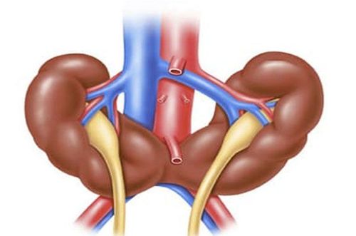
Normally, the kidneys are located in the renal fossa on both sides of the spine and are separate from each other, in horseshoe nephropathy the left and right kidneys are connected by a false isthmus, in more than 90% of cases the adhesions are at the lower pole of the kidney. . Call the isthmus pseudorenal because this contact site is very small, difficult to detect and this isthmus has no function. When the two kidneys are joined together, they are shaped like a horseshoe, so it is called horseshoe kidney disease.The formation of horseshoe kidney is caused by a certain cause that affects the development of the kidneys and changes position, they will develop in the pelvic region and gradually move to the sides of the lumbar spine. When moving the upper pole, the kidney rotates inward so that the kidney is directed into the spine. This process of growth and migration is completed before 8 weeks of gestation.
Currently, the cause of horseshoe kidney malformation is not clearly known. It has been found that the incidence of horseshoe kidney disease in men is twice as high as in women. Usually the disease does not cause any symptoms, but complications can cause unusual symptoms.
Complications of horseshoe kidney include:
Horseshoe kidney can cause complications such as:
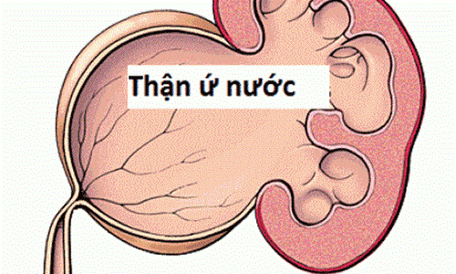
Urinary tract obstruction, urinary retention: Patients feel tension in the pelvic area, little urine, urine retention for a long time. urinary infection. Formation of kidney stones with a rate of about 20-60%: The patient feels sharp pain in the back, lower abdomen, urine with sediment. Urinary tract infection: Fever, cloudy urine, pus or blood, painful urination, frequent urination, discomfort when urinating. Kidney cancer: It has been found that up to 45% of renal carcinoma cases are seen in patients with horseshoe kidney. Part of the reason is that the effects of hydronephrosis, kidney stones and urinary tract infections increase the risk of kidney cancer in these patients. Due to abnormalities in position and anatomical shape, patients with horseshoe kidney are at increased risk of kidney damage during trauma.

2. Ultrasound image of horseshoe kidney
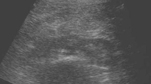
Because the disease often has no clinical manifestations, it is often difficult for patients to detect horseshoe kidney disease. Therefore, patients are often discovered by chance during examination, ultrasound is the method that can see this malformation and sometimes show images of complications caused by horseshoe kidney.Ultrasound is a test that uses ultrasound waves to create pictures of organs in the body. Renal ultrasonography allows assessment of structural abnormalities, focal or diffuse renal injury.
Ultrasound images that can be seen on horseshoe kidney patients include:
Ultrasound shows the kidneys are out of alignment with normal. That is, normally the upper pole of the kidney will point towards the spine. But in horseshoe nephropathy, the lower pole of the kidney is seen towards the spine. However, to detect this, the sonographer needs experience and careful investigation. Detecting pseudo-renal isthmus: When detecting a kidney that is off-axis compared to normal, the doctor will find the junction of the two kidneys to diagnose horseshoe kidney. But in patients with large body, thick abdominal fat layer, then observing the horseshoe kidney will be very difficult at that time. Detect complications caused by horseshoe kidney such as: See the dilatation of the renal calyces, hydronephrosis and pus stasis. Kidney stone: An echogenic structure, the size is not fixed, it can be large or small, accompanied by echogenic shadow behind. Detecting an abnormal tumor in the kidney. Ultrasound is sometimes limited, sometimes overlooked, and often difficult to distinguish between a horseshoe kidney and an ectopic kidney. It takes an experienced sonographer and a good system of machines for such a good ultrasound to limit the chance of missing an injury during abdominal ultrasound.
If the ultrasound results are suspicious, the doctor will order other tests to help accurately diagnose the disease. Taking UIV, CT-scan or magnetic resonance imaging to help diagnose the disease accurately, detect other abnormalities on the kidney.

3. Notes on horseshoe kidney
When detecting horseshoe kidney disease, the patient should pay attention to the following issues:Horseshoe kidney is an abnormal condition, however, it does not mean that every horseshoe kidney must be treated. There are some people who have horseshoe kidney but it does not cause any harmful signs or complications to the body. So just follow up periodically. There is no specific treatment for people with horseshoe kidney disease. However, when suffering from the disease, the patient should regularly undergo regular check-ups to detect early complications caused by the disease and treat it early. Patients are treated when having complications of the disease, which can be medical treatment or surgical treatment depending on each specific complication. Kidney injury is more common with abdominal trauma in people with horseshoe kidney. Therefore, it is necessary to pay more attention and if there is a minor abdominal injury, it should be checked. When detecting abnormal symptoms of the urinary system, it is advisable to examine for early detection of urinary tract disease and to detect or exclude horseshoe kidney. Periodic examination is the best way to detect horseshoe kidney disease early, when detected, regular monitoring is required to detect early complications caused by horseshoe kidney.
Vinmec International General Hospital with a system of modern facilities, medical equipment and a team of experts and doctors with many years of experience in neurological examination and treatment, patients can completely peace of mind for examination and treatment at the Hospital.
If you have a need for medical examination at Vimec Health System nationwide, please make an appointment on the website to be served.
Please dial HOTLINE for more information or register for an appointment HERE. Download MyVinmec app to make appointments faster and to manage your bookings easily.


