
This is an automatically translated article.
The article is professionally consulted by Master, Doctor Trinh Thi Phuong Nga - Radiologist - Department of Diagnostic Imaging and Nuclear Medicine, Vinmec Times City International Hospital.Pulmonary infarction is a dangerous disease with a high risk of death if not detected and treated promptly. Accurate diagnosis of pulmonary infarction is one of the measures to help doctors intervene early and get positive results for patients.
1. What is a pulmonary infarction?
Pulmonary infarction is also known as pulmonary embolism. This is a condition in which a blood clot enters a blood vessel in the lungs, blocking the normal flow of blood. This blockage interferes with air exchange. Depending on the size of the clot and the number of blood vessels involved, a pulmonary infarction can be life-threatening.Most blood clots that cause pulmonary embolism are formed in the veins of the legs. In addition, blood clots can also originate from veins in the pelvis, trunk, upper extremities, or from the right heart. Some cases of pulmonary infarction are not caused by blood, but by fat embolism (with fat floating in the blood vessels) or by foreign bodies, cancer, intravascular air, amniotic fluid,...
Symptoms of pulmonary infarction often occurs suddenly, with manifestations including:
Shortness of breath and rapid breathing; Chest pain; Hemoptisi ; heart palpitations; May faint. SEE ALSO: Role of electrocardiogram in the diagnosis of pulmonary infarction
2. Medical techniques to diagnose pulmonary infarction
Most patients with pulmonary infarction have vague symptoms with varying degrees of severity, and investigations should be performed to exclude or confirm the diagnosis. The combination of clinical symptoms, risk factors, and medical techniques makes a fairly accurate assessment of the likelihood of pulmonary infarction. The diagnostic techniques include:Electrocardiogram: Only 6% of patients with acute major pulmonary embolism and 23% of patients with subacute pulmonary embolism have normal ECG. If the pulmonary embolism is small and hemodynamically stable, only sinus tachycardia is usually present. Cardiac arrhythmias such as acute atrioventricular block or ventricular arrhythmias are rare. The most common finding is nonspecific ST and T-segment changes, which are often transient. The image of acute bronchiectasis accounts for only about 30% of cases of large-scale pulmonary embolism. Use an electrocardiogram primarily to rule out other problems such as pericarditis or myocardial infarction.
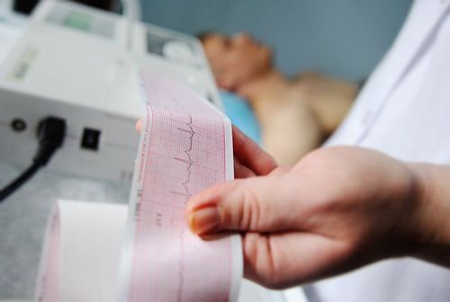
Điện tâm đồ là một trong nhiều phương pháp chẩn đoán nhồi máu phổi
Blood tests: Pulmonary infarction cases often have elevated LDH, increased indirect bilirubin and normal SGOT. The patient also had a slight increase in white blood cells (12,000-13,000/ml) and a slight decrease in hematocrit (30-35%), and a slight decrease in platelets (100,000-200,000/ml) even in the absence of complications from taking heparin. The fibrin degradation product of fibrin containing D-dimer was increased by more than 500 ng/ml in more than 90% of patients with pulmonary infarction; Arterial blood gases: Patients with pulmonary infarction often have decreased PaO2 and normal or decreased PaCO2 (due to hyperventilation). Extensive pulmonary embolism can cause respiratory and metabolic acidosis. About 14-38% of patients with pulmonary infarction still have normal arterial blood gas results. PaO2 is normal in small pulmonary embolism and abnormal in large pulmonary embolism. Increased physiological dead space ventilation is a very sensitive indicator for the diagnosis of pulmonary infarction, closely related to the degree of pulmonary embolism and disease progression; Transthoracic or esophageal echocardiography: Helps doctors evaluate right ventricular hypertrophy, detect heart chamber clots and rule out other conditions such as hypovolemic shock, aortic dissection, disease pericardial, valvular, myocardial infarction,... Echocardiography is considered an auxiliary tool (not the main diagnostic tool) in determining pulmonary infarction pathology; Pulmonary scintigraphy: It is an indirect means of diagnosing pulmonary infarction because it cannot detect blood clots, but only detects the resultant areas of impaired perfusion. A normal pulmonary perfusion scan helps to rule out a diagnosis of pulmonary embolism. The ability to diagnose pulmonary infarction of this technique is divided into 3 categories: high - moderate - low. High probability imaging allows 85% correct diagnosis. However, the majority of patients with pulmonary infarction only obtain moderate or low perfusion scintigraphy. This technique plays an important role in deciding the treatment direction for the patient; Computed tomography: Includes normal CT, spiral CT (multi-sequence) and electron beam CT. Spiral CT scan using intravenous contrast or electron beam CT is a non-invasive diagnostic method with many advantages. In many places, helical CT scan is the first measure for diagnosis because the imaging technique is fast, uncomplicated, can detect right ventricular dilatation compared with the traditional method of pulmonary angiography, and at the same time gives a clear and accurate image. more complete (both vascular and parenchymal) than radiographic imaging. On CT scan, the manifestation of pulmonary infarction is very obvious in about 90% of cases; Nuclear magnetic resonance imaging (MRI): A non-invasive imaging method, allowing doctors to accurately assess the anatomical morphology - function of pulmonary perfusion and right ventricular function. The advantages of MRI compared to CT and pulmonary angiography are: Avoiding contrast-induced nephrotoxicity, angiography within 1 stroke, high sensitivity and specificity in detecting deep vein thrombosis and thrombosis. mass in the pulmonary artery. However, the prolonged time of MRI (15 - 30 minutes) should cause many disadvantages for the group of patients with unstable hemodynamics; Pulmonary angiography: Selective pulmonary angiography is considered the gold standard in diagnosing or excluding pulmonary infarction. The doctor will order pulmonary angiography when there is hypotension, collapse or the patient cannot be diagnosed by other means. This technique is relatively contraindicated in patients with pregnancy, high bleeding risk, renal failure, and blood clots in the right heart. Cannography can be done through the femoral vein, brachial vein, internal jugular vein, and subclavian vein. Before taking cardiac catheterization, it is necessary to perform cardiac catheterization to assess cardiac output, shunt, degree of hemodynamic disturbance,... Pulmonary angiography is mandatory if measures to take or dilate thrombus by catheter are applied.

Chụp MRI là phương pháp chẩn đoán nhồi máu phổi không xâm lấn, nhiều uwuu điểm
The above medical techniques are indicated to perform the diagnosis of pulmonary infarction. The method that is used and brings the highest diagnostic efficiency should be applied is the CT scan with intravenous contrast injection. From there, the doctor will have an accurate conclusion and make an appropriate treatment intervention plan for the patient. However, for accurate diagnosis results, patients should choose reputable medical facilities to perform.
Currently, Vinmec International General Hospital has been and continues to be fully equipped with modern diagnostic facilities such as: PET/CT, SPECT/CT, MRI, electrocardiogram..., blood tests, Ultrasound to perform examination and treatment of diseases, including pulmonary infarction. The entire examination process at Vinmec is performed by a team of experienced and qualified medical professionals, so the results are accurate, and the patient has the opportunity to receive early treatment to ensure long-term health.
Please dial HOTLINE for more information or register for an appointment HERE. Download MyVinmec app to make appointments faster and to manage your bookings easily.