This is an automatically translated article.
The article was professionally consulted by Specialist Doctor I Nguyen Thanh Hai - Radiologist - Department of Diagnostic Imaging and Nuclear Medicine - Vinmec Times City International General Hospital. Doctor Hai has more than 20 years of experience in the field of diagnostic imaging, especially in the field of multi-slice computed tomography, magnetic resonance.1. Overview of fatty liver and cirrhosis
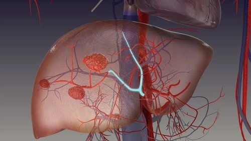
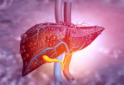
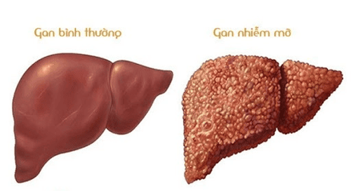
Fatty liver is a condition in which 5-6% more fat accumulates in the liver. Although it does not directly affect life, but if the patient is subjective, without treatment, a long-term fatty liver can lead to the death of liver cells, the formation of scar tissue to replace healthy liver cells. , reducing blood flow to the liver. Fatty liver disease can be complicated into cirrhosis, liver failure, liver cancer, seriously affecting the patient's health.There are two main causes of fatty liver, they are:
Alcoholic fatty liver disease (AFLD): occurs due to the patient drinking too much alcohol. Nonalcoholic fatty liver disease (NAFLD): occurs when a person's body has an abnormal metabolic state. NAFLD has been linked to obesity, high cholesterol, and diabetes. This is the most common form of fatty liver in developed countries. Some other causes can also cause fatty liver such as viral hepatitis, use of certain drugs (such as aspirin, steroids, tamoxifen, tetracycline), pregnancy,...
Because there are very few symptoms and signs The symptoms are clear, so most patients with fatty liver and cirrhosis in the early stages do not know they have the disease. Some people may experience mild fatigue or abdominal discomfort. As the disease progresses, patients will experience many symptoms such as loss of appetite, nausea, fatigue, weight loss, yellowing of the skin and eyes, swelling in the legs and abdomen,...
2. How to diagnose fatty liver disease and cirrhosis?
To diagnose fatty liver disease and cirrhosis, your doctor will take a patient's history and symptoms. Some tests such as blood lipid test, AST test, ALT test will be done to evaluate the amount of fat in the blood and liver function.In addition, to diagnose fatty liver disease and cirrhosis accurately, the doctor will only perform one or more imaging techniques such as:
2.1. Liver ultrasound To perform an ultrasound of the liver, the doctor will use an ultrasound machine with an instrument that sends ultrasound waves to the upper right side of the patient's abdomen to look at the image of the liver. The images will be displayed on the screen, helping the doctor to detect abnormalities in the liver structure and damage to the liver.
If the patient has a fatty liver, the liver ultrasound images will appear scattered or concentrated in areas. Fatty liver looks brighter than normal liver. In addition, fatty liver can be diagnosed if the echogenicity of the liver is superior to that of the renal cortex and spleen, there is also impaired echocardiography, loss of diaphragm clarity, and poor delineation of the Intrahepatic structures
If the patient has cirrhosis, the ultrasound image will have small nodules on the liver surface, liver margins convex, many large nodules, peritoneal fluid and rough parenchyma, tail lobe hypertrophy, atrophy right lobe, ... Based on the abnormality of the liver on ultrasound, the doctor will assess the degree of cirrhosis.
2.2. Computed tomography (CT) of the liver Computerized tomography is a technique that combines X-ray technology and a computer system to analyze images through sections of X-rays. Computed tomography scans give more detailed images, helping to evaluate cirrhosis, fatty liver more accurately than conventional ultrasound.
If the liver is normal, on computed tomography without contrast, the liver will have a much larger density than the spleen and blood, the blood vessels in the liver can be seen as structures relatively reduced proportion. Fatty liver disease is diagnosed when the density of the liver is at least 10 HU below that of the spleen or if the density of the liver is less than 40 HU. If the fatty liver is severe, the blood vessels in the liver may be hyperattenuated compared with fatty liver tissue.
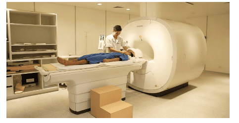
2.3. Magnetic resonance imaging (MRI) in the evaluation of cirrhosis and fatty liver Magnetic resonance imaging uses a magnetic field and radio waves to create detailed images of the liver. Magnetic resonance images with high contrast, clear, sharp, good anatomical properties, serve well for diagnosis and treatment. For fatty liver, magnetic resonance imaging is the most sensitive fatty liver imaging technique today, with very high accuracy even in mild fatty liver. Some MRI scanners can also calculate the percentage of fat in the liver.
MRE (MR elastography) is a special MRI technique used to evaluate cirrhosis. The movement of small vibrational waves in the liver is captured by the machine to create a visual map of elasticity or stiffness throughout the liver, helping to detect cirrhosis at an early stage.
2.4. Elastic ultrasound Elastography is a special ultrasound technique that helps to evaluate the stiffness of tissue through the degree of elasticity of the tissue when subjected to mechanical force. Liver stiffness is related to the degree of liver fibrosis. Elastography helps to assess liver fibrosis quickly without the need for invasive procedures. This is an effective technique in the assessment of cirrhosis, helping to screen and detect it at an early stage.
In addition, in some cases, to evaluate cirrhosis and fatty liver, the doctor may order a liver biopsy. Under imaging guidance, a fine needle is inserted to obtain a small sample of liver tissue. The specimen will be examined under a microscope for signs of steatosis, inflammation, damage, and fibrosis.

To detect fatty liver disease and cirrhosis at the earliest, we need to have regular health check-ups, with tests, imaging and clinical examinations performed by doctors, which will help patients easily detect fatty liver from the very first stage.
Vinmec International General Hospital is a high-quality medical facility in Vietnam with a team of highly qualified medical professionals, well-trained, domestic and foreign, and experienced.
A system of modern and advanced medical equipment, possessing many of the best machines in the world, helping to detect many difficult and dangerous diseases in a short time, supporting the diagnosis and treatment of doctors the most effective. The hospital space is designed according to 5-star hotel standards, giving patients comfort, friendliness and peace of mind.
If you have a need for consultation and examination at the Vinmec Health System Hospitals nationwide, please book an appointment on the website for service.
Please dial HOTLINE for more information or register for an appointment HERE. Download MyVinmec app to make appointments faster and to manage your bookings easily.
Reference source: radiologyinfo.org


