This is an automatically translated article.
The article is professionally consulted by Master, Doctor Nguyen Huy Nhat - Department of Medical Examination & Internal Medicine - Vinmec International General Hospital Da Nang.ECMO is indicated in cases of respiratory failure or acute circulatory failure. ECMO works to replace the activity of the heart - lungs so that the patient has time to recover and the doctor also has time to find the cause to help treat the disease effectively.
1. What is acute respiratory failure?
Acute respiratory failure is a condition in which the lungs suddenly fail to respond to the body's gas exchange function, resulting in hypoxia with or without hypercapnia. Depending on the individual case, hypoxemia alone can be milder or more severe than hypoxia with hypercapnia, especially in adult respiratory distress syndrome (ARDS). .The risk of acute respiratory failure is greater for people with pneumonia, sepsis, aspiration of gastric juice, and severe trauma. Severe respiratory failure in adults characterized by pulmonary edema with increased permeability, severe arterial hypoxia, and decreased carbon dioxide emissions. The disease is treated by various methods such as mechanical ventilation, intravenous fluids, prevention of venous thrombosis, especially lung-protective mechanical ventilation with low tidal volume and the new ECMO technique. Drug treatment has not yet yielded significant results.
2. Symptoms of acute respiratory failure
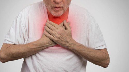
Khó thở là triệu chứng thường thấy của suy hô hấp cấp
Pale, cyanotic skin: Often there is cyanosis in the lips and extremities when the reduced Hb is above 5g/100ml, SaO2 is below 85g/l. The skin turns red, sweaty when PaCO2 increases as much as in an acute episode of chronic bronchitis.
Cardiac arrhythmia: Patients can be diagnosed with sinus tachycardia, tachycardia (atrial fibrillation, flutter), ventricular fibrillation as the final manifestation. The most dangerous is the case of severe hypoxia or the PaCO2 is too low, which can cause cardiac arrest if not intervened within 5 minutes of the phenomenon.
Blood pressure increases or decreases : In the first stage, blood pressure is usually high, then gradually decreases. In these cases, immediate intervention is required by means of balloon squeezing, endotracheal intubation, sputum suction, and mechanical ventilation.
Nervous and Consciousness Disorders : The brain consumes 1/5 of the total oxygen of the whole body. So it is the organ that suffers the earliest consequences of hypoxia and hypercapnia. Patients may have neurological disorders with manifestations of struggling, confusion and loss of tendon reflexes. Or there are manifestations of consciousness disorders such as lethargy, coma.
Lung problems: Auscultation of the lungs with moist rales, sometimes with atelectasis, respiratory paralysis (intercostal muscle palsy, epipharyngeal paralysis), pleural effusion seen in mechanically ventilated patients or after placement of a lower venous catheter stroke, bronchopneumonia in the posterior region of the lungs.
3. Diagnosis of acute respiratory failure
Diagnosis of acute respiratory failure must be based on many factors, including:Shortness of breath, cyanosis of the lips and extremities Poor response to oxygen therapy Chest X-ray: X-ray film showing lesions Bilateral diffuse alveolar worsening. Arterial blood gas: Blood gas test showed decreased blood oxygen, PaO2/FiO2 ≤ 300 mmHg with PEEP ≥ 5 cmH2O. Because the symptoms of acute respiratory failure are similar to some other diseases, it is necessary to make a differential diagnosis such as acute pulmonary edema, renal artery stenosis, head trauma, pulmonary venous obstruction, pulmonary vasculitis. , acute pneumonia.
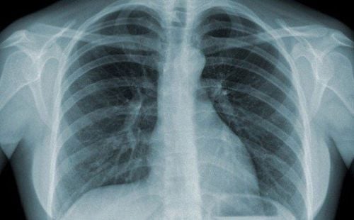
X-quang ngực là một trong những yếu tố để chẩn đoán suy hô hấp cấp
4. Treatment of acute respiratory failure in adults
Pleural drainagePleural drainage is indicated in cases of pneumothorax, pneumomediastinum and in hemothorax, pleural effusion. In some serious cases such as bronchial rupture and tear, in addition to draining the patient, there is the possibility of surgical intervention and intubation of a Carlens tube or endotracheal tube. Pleural drainage may also be indicated prophylactically in patients with multiple rib fractures and mobile rib plaques prior to endotracheal anesthesia or mechanical ventilation. The drain is removed within 48-72 hours after the air or fluid is gone.
Open airway
Open airway is the first job to do for patients with acute respiratory failure. Depending on the cause and degree of difficulty in breathing, the following procedures are applied:
Hook mouth, nose, throat, suck out dirt, sand, mud, food, blood, then wipe it off Lifting jaw, put on Mayo canuyn to lift tongue, put the head very back or pull the tongue out when the tongue is retracted. Threading a polyethylene wire through the thyroid membrane: This method is applied to patients with extensive sputum obstruction that cannot be aspirated in time and cannot be completely drained. This method is simple to perform and in most cases, after only 24 - 48 hours, the 2 lungs have been cleared and the wire removed. Widely aspirate sputum, blood in the tracheobronchial tubes by suction or suction with a bronchoscope or use a laryngoscope to lift the epiglottis, expose the larynx, insert the suction tube deep into the trachea and aspirate or use a bronchoscope - bronchi and suction (bronchoscopies aspiration). Intubation, tracheostomy Water and fluid balance in the body
Reducing left atrial filling pressure by limiting fluid and using diuretics is important in the treatment of ARDS. Maintaining a normal or low left atrial filling pressure minimizes pulmonary edema, prevents arterial hypoxia and lung elasticity, shortens the duration of mechanical ventilation and hospital stay, and reduces morbidity and mortality. mortality in ICU and surgical patients. Extra care should be taken in patients with hemodynamic instability and hypoperfusion of vital organs such as the kidney.
Medication
Corticosteroids: Corticosteroids work to reduce inflammation in the lungs. The drug is not recommended to be used in high doses. However, there are specific situations where corticosteroids can be used with caution when the underlying cause responds to corticosteroid treatment such as idiopathic organizing pneumonia. Antibiotics: in case of signs of infection Diuretics: congestive heart failure, acute hemodynamic pulmonary edema, volume overload Plasma exchange to remove antibodies in autoimmune diseases causing respiratory muscle paralysis such as myasthenia gravis chance, Guillain-Bare syndrome.

Có thể điều trị suy hô hấp cấp ở người lớn bằng thuốc
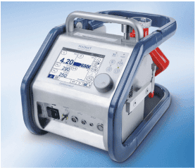
5. Treatment of severe respiratory failure in adults with ECMO
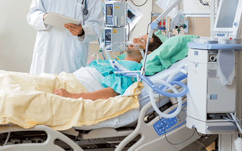
The ECMO system has the potential to replace the function of the heart and lungs. People who need support from an ECMO machine need intensive care (ICU) in a hospital. Usually, people who need support only use the ECMO machine for a few hours to a few days, but some people need to use ECMO for a few weeks, depending on the progression of the disease. ECMO is indicated in the following cases:The lungs cannot supply enough oxygen (O2) to the body even with oxygen The lungs cannot eliminate carbon dioxide (CO2) even with help from a ventilator Heart Inability to pump enough blood to meet the body's needs In general, adult ECMO is indicated for patients with potentially reversible acute respiratory failure, acute circulatory failure, unresponsive to routine treatment. Deploying ECMO technique for patients needs to comply with indications and contraindications.
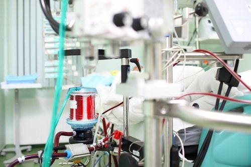
The ECMO machine is connected to the patient through plastic tubes (cannula tubes) that are inserted into large veins in the legs, neck or chest. The ECMO machine pumps blood from the patient's body to an artificial lung (oxygenator) to replenish O2 and remove CO2 from the blood, then put it back into the bloodstream. So it will replace the patient's cardiopulmonary function.
Before the procedure, the doctor will order a blood gas test, use anticoagulants, some sedatives, and anesthesia.
After placing the ECMO machine, possible complications include bleeding due to the effects of anticoagulants, kidney failure when blood is not fully supplied to the kidneys, infections due to long-term treatment while the system is not functioning properly. The machine system is in direct contact with the patient's body, leg damage because the system is connected to the body through the femoral artery, cerebral stroke due to insufficient blood supply to the brain when blood clots appear.
ECMO technique is being successfully applied in the treatment of circulatory failure and acute respiratory failure at Vinmec International General Hospital. This is a difficult medical technique, requiring doctors and technicians to have high professional expertise.
Besides, the hospital has invested in modern machinery and equipment to support doctors during surgery and patient resuscitation, such as: Maquet's HR20 artificial heart and lung system, Machine breathe GE's R860.
Please dial HOTLINE for more information or register for an appointment HERE. Download MyVinmec app to make appointments faster and to manage your bookings easily.