This is an automatically translated article.
Hydronephrosis due to ureteral pyelonephritis is an internal or external narrowing of the junction between the renal pelvis and the ureter. Treatment of hydronephrosis due to narrowing of the ureteropelvic junction follows the principle of damage resolution and prevention of complications.1. Hydronephrosis
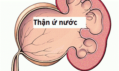
Hydronephrosis due to narrowing of the pyelonephritis - ureteral junction is the impairment of urine flow from the renal pelvis to the ureter, causing dilatation of the collection system and potentially impaired renal function. Dilation of the kidney can cause compression of the renal parenchyma leading to decreased renal filtration or infection causing kidney damage.Narrow pyelonephritis - ureteral junction is the most common cause of obstructive disease in children, most commonly around 5 years of age. The prevalence of hydronephrosis due to narrowing of the pyelonephritis - ureteral junction in men is twice that of women. Hydronephrosis on the left is more common than on the right, and hydronephrosis on both sides accounts for about 10%.
2. Diagnosis
To diagnose hydronephrosis due to narrowing of the ureteropelvic junction, it is necessary to conduct a clinical examination, and do additional paraclinical tests including:Tests: urea, complete blood count, urinalysis, creatinine,... Ultrasound: shows the size of the kidney, the thickness of the renal parenchyma and also helps to distinguish it from dilatation of the ureter, detecting polycystic kidney on the other side. X-ray: intravenous contrast urography, bladder contrast during urination, kidney scan with Tc99m. In addition, it is necessary to make a differential diagnosis for the following cases:
Hydronephrosis due to ureteral aneurysm: differentiate based on ultrasound Hydronephrosis due to vesico-ureteral reflux Double kidney Polycystic kidney Case of dumb kidney on X-ray- X-ray: Ultrasound will help confirm this is a hydronephrosis due to narrowing of the junction or due to dilatation of the ureter. If polycystic kidney, ultrasonography confirmed no damage to the renal pelvis and calyces.
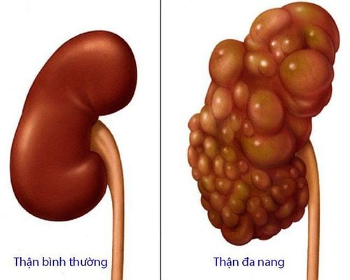
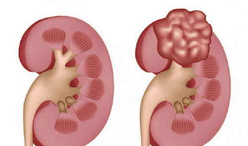
Bệnh thận ứ nước cần được chẩn đoán phân biệt với bệnh thận đa nang, tránh điều trị sai hướng
3. Treatment of hydronephrosis
There are many methods of treating hydronephrosis due to narrowing of the ureteropelvic junction, depending on each case, the doctor will give an appropriate treatment method. However, the methods still follow the principle of resolving damage and preventing complications caused by hydronephrosis.3.1 Conservative treatment Indications for conservative treatment: when surgery is not indicated. Conservative treatment:
Monitor the size of the kidney - pyelonephritis on ultrasound every 3 - 6 months. If the kidney is enlarged, reassess renal function on renal scintigraphy. Antibiotics for UTIs. Prophylactic antibiotics: severe bilateral hydronephrosis, hydronephrosis detected before birth with RPD > 15mm. Drug of choice: amoxicillin 12-25mg/kg/day orally. The duration of drug use starts immediately after birth until the combined urinary system abnormalities such as vesicoureteral reflux, posterior urethral valve are excluded. 3.2 Surgical treatment 3.2.1 Indications Surgery is indicated when:
There are symptoms: abdominal pain, infection, kidney stones, palpable large kidney. Asymptomatic but with reduced renal function less than 40% on renal scan. Failure of conservative treatment: increased renal size on ultrasound and renal function less than 40% or decreased renal function more than 10% on scan compared with previous. Giant hydronephrosis with anteroposterior renal pelvis diameter – RPD > 50mm on ultrasound. 3.2.2 Surgical methods The surgical steps include:
Shaping the pyelonephritis - ureter according to Anderson - Hynes through laparoscopic, open surgery or robotic surgery. Urinary catheterization with a JJ catheter or a ureteral stent or a percutaneous nephrostomy catheter. When renal function is less than 10% on scintigraphy: radical surgery or pyelonephritis. Renal drainage: After 1-2 months of drainage, renal function will be re-evaluated. If kidney function improves, pyeloplasty is performed. Nephrectomy for the following cases: Complete loss of kidney function. After pyelonephritis, renal function did not improve. Small atrophic hydronephrosis with loss of function or recurrent urinary tract infections. After surgery, patients need to be treated with antibiotics, and pain relief after surgery. Place a ureteral stent and remove it 5-7 days after surgery. For JJ catheterized patients, withdraw the JJ through cystoscopy after 1-2 months.

Bệnh nhân sẽ được chỉ định cắt thận nếu như chức năng thận mất hoàn toàn
4. Common Complications
After surgery, the patient may experience some complications such as:Bleeding Infection Anastomal fistula Obstruction anastomosis Complications related to JJ Catheter Obstruction Recurrent anastomosis Therefore, after surgery Patients should be closely monitored, with ultrasound 1 month after surgery and every 3-6 months for at least 2 years to assess the improvement of fluid retention and symptoms. If 3-6 months after surgery, the degree of fluid retention does not change or worsens or renal scintigraphy is indicated to re-evaluate the surgical results,
In summary, hydronephrosis due to stricture of the ureteropelvic junction is an internal or external narrowing of the junction between the renal pelvis and the ureter. Treatment of hydronephrosis due to narrowing of the ureteropelvic junction can be treated conservatively or surgically when indicated. After surgery, the patient may experience a number of complications, so when there are any abnormal signs, it is necessary to immediately notify the medical staff, periodically visit for timely treatment.
Vinmec International General Hospital is a high-quality medical facility in Vietnam with a team of highly qualified medical professionals, well-trained, domestic and foreign, and experienced.
A system of modern and advanced medical equipment, possessing many of the best machines in the world, helping to detect many difficult and dangerous diseases in a short time, supporting the diagnosis and treatment of doctors the most effective. The hospital space is designed according to 5-star hotel standards, giving patients comfort, friendliness and peace of mind.
Please dial HOTLINE for more information or register for an appointment HERE. Download MyVinmec app to make appointments faster and to manage your bookings easily.


