This is an automatically translated article.
The article is professionally consulted by Master, Doctor Tong Diu Huong - Radiologist - Department of Diagnostic Imaging - Vinmec Nha Trang International General Hospital.Colon cancer is a fairly common disease in our country, usually in adult men. This is also the cause of intestinal obstruction. The disease needs to be detected early for surgical intervention.
1. Overview
1.1. Stenosis of the colon
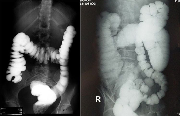
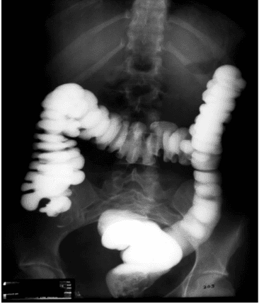
Colorectal cancer is a very common disease today, not only in developed countries but also in developing countries, including Vietnam. In general, there are 3 types of colon cancer, including those that cause narrowing of the colon, those that grow in the colon wall, and those that grow in the colon wall. The type of cancer that causes obstruction of the colon is most often found at the junction between the sigmoid colon and the rectum. The lesion is a small tumor, which causes the lumen of the colon to narrow. Cancerous tumors in the right colon rarely narrow the bowel, whereas tumors in the left colon, although small, tend to narrow the colonic lumen.1.2. Treatment of intestinal obstruction
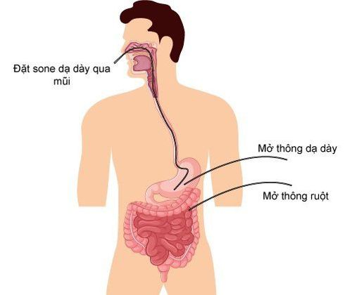
The purpose of colonic stenting before surgery is to clean this part before removing the tumor and connecting the colon, reducing the patient's need for 2 surgeries.For a long time, flexible endoscopic technique has played an important role when wanting to intervene in the colon before and after surgery. However, there are also many cases where flexible bronchoscopy fails because it cannot pass through the narrow site of obstruction.
Meanwhile, interventional radiology is minimally invasive and uses smaller sized instruments than flexible endoscopes. Therefore, this technique can overcome narrow areas that flexible bronchoscopy cannot. This is considered an additional option in clinical practice.
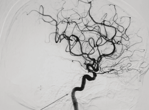
Digital angiography with background erasure is an imaging method that combines X-ray and digital processing. The images obtained before and after injecting contrast material into the patient's body are processed to remove the background, displayed fully and clearly.

2. Indications and contraindications
Treatment of colonic occlusion under the guidance of digital angiography erasing background is indicated in the following cases:Preparing for surgery for colon cancer 1 stage; Narrowing of the colonic anastomosis after surgery; Primary or secondary colon cancer but no longer indicated for radical surgery; Stenosis of the colon is rare: Due to tuberculosis, diverticulitis, colitis after irradiation or after colonic fistula. In contrast, contraindicated for people with perforated hollow organs or systemic infections.
3. Preparation steps
3.1. Executor
The team responsible for conducting digital angiography to erase the background to treat colonic stenosis includes:Specialist doctors and supporting medical staff; Electro-optical technician; Nurses; Anesthesia team if the patient cannot cooperate.
3.2. Vehicle
Digital Subtraction Angiography (DSA); Film, printers and image storage systems; Lead vest and apron for X-ray shielding.3.3. Medicine
Local anesthetic or general anesthetic if indicated; Water-soluble Iodine Contrast; Medicines to limit spasms of the digestive tract; Antiseptic solution for mucous membranes and skin.3.4. General medical supplies
Syringes of sizes 5, 10, and 20ml; Distilled water / physiological saline ; Full surgical protective suit; Sterile interventions: Knives, scissors, tongs, metal bowls, bean trays and tool holders; Cotton swabs, gauze and surgical tapes; Medicine box and emergency equipment if there is a contrast agent accident.3.5. Special medical supplies
Kim Chiba soft tissue aspiration; Set of conduits into the lumen; Standard conductor 0.035 inch; Hard conductor 0.035 inch, length 260 - 300cm; Cobra 4-5F angiography catheter; Balloons; Pressure pump; The bracket (also called a stent).3.6. Patient
The procedure is carefully explained to coordinate well with the doctor; Fasting before 6 hours (Otherwise, you can drink less than 50ml of water); In the intervention room: The patient lies on his back, has a machine to monitor blood pressure, breathing rate, electrocardiogram, SpO2 and pulse; Cover with a sterile towel with holes; If the patient does not lie still, too excited, need to give sedation...3.7. Test form
Medical records for inpatient treatment; The order form for the procedure has been approved; X-ray film, CT scan, MRI (if any).
4. Procedure
4.1. Narrow location review
Insert the catheter and guide through the anus into the colon to reach the stricture; Withdraw the lead, then inject a water-soluble contrast agent to help assess the extent and location of the stenosis.4.2. Approaching a narrow location
Continue to thread the guide wire through the narrow position under the guidance of the enhanced X-ray screen; Pass the conduit through the narrow passage by the conductor; Inject the contrast agent through the catheter to determine the extent, location, and length of the narrowing.4.3. Spread and place the stand
Insert the rigid conductor into the conduit through the narrowing; Use a narrow-gauge balloon over a rigid conductor; Place and release the stent holder through the rigid lead.4.4. End of the trick
Use contrast agents to check circulation from the stomach to the duodenum and jejunum; Withdraw all leads and catheters.5. Evaluation of results
About the position: Place the stent in the correct position of the stenosis, the upper and lower ends of the stent cover the 2 ends of the stenosis at least 1cm; Function: Drugs pumped from upstream have circulation to downstream. The lumen of the alimentary canal is not more than 30% narrow; Do not drain the contrast agent out of the gastrointestinal tract into the abdomen or retroperitoneal space.6. Complications and treatment
Stent slip: The cause is the choice of stent size is not suitable for the narrowing; Intestinal obstruction: Stent sliding downstream or fibrous, undercooked food stuck to the stent; Hollow visceral perforation: Immediate emergency surgery is required; Gastrointestinal bleeding: Continue monitoring and medical treatment. Embolization can be treated if the bleeding does not stop on its own. Thanks to the DSA background digitized angiogram system, many diseases have been clearly diagnosed and promptly intervened, including treatment of colonic stenosis. For colon cancer, for effective surgical intervention, only 1 surgery should be performed. If all patients arrive late to the hospital, there is a risk of not being able to remove the cancerous tumor, leading to many unfortunate consequences.In order to improve the medical examination and treatment process, Vinmec International General Hospital has been providing a system of modern and standard medical machines for examination, treatment and prevention. many diseases. In particular, to meet the requirements of using comprehensive medical services, Vinmec International Hospital is also implementing a Package of Screening and Early Detection of Colorectal Cancer, implemented by a team of doctors and nurses. industry leader with rich experience in the field of diagnosis and treatment of colorectal cancer. When using this service package, you will have the support of a system of modern standard equipment and technology, full of specialized facilities for diagnosing diseases and staging pre-treatment stages such as: CT scan , Endoscopy, PET-CT scan, MRI, Mammogram, histopathological diagnosis, gene-cell testing,... help detect colon cancer early even when there are no warning symptoms. brand.
If you have a need for consultation and examination at Vinmec Hospitals under the nationwide health system, please book an appointment on the website for service.
Please dial HOTLINE for more information or register for an appointment HERE. Download MyVinmec app to make appointments faster and to manage your bookings easily.


