This is an automatically translated article.
This article was written by Specialist Doctor II Khong Tien Dat, Doctor of Radiology - Department of Diagnostic Imaging - Vinmec Ha Long International Hospital.Transcranial doppler ultrasound is a painless, noninvasive ultrasound technique that uses high-frequency sound waves to measure the speed and direction of blood flow in the blood vessels of the brain. This test is intended to check and record the rate of blood flow in the arteries of the brain, facilitating the diagnosis and screening of diseases that may affect this organ.
1. What is transcranial doppler ultrasound?
Transcranial doppler ultrasound is a non-invasive diagnostic tool that uses ultrasound waves to measure blood flow in the brain. The brain is considered the control center of the body's activities. Although the function of the brain depends on other organs, the brain still plays a role in controlling most activities. It is the millions of cells and nerves that are connected to other organs that dictate the actions necessary to continue life.Due to intense metabolic function, the brain requires at least 20% of oxygen coming from the heart through a tight network of blood vessels. If too much blood reaches the brain, increased intracranial pressure occurs and can lead to tissue damage. Conversely, if the blood flow to the brain is reduced, the brain parenchyma is not supplied with enough nutrients, becomes dysfunctional and necrotic.
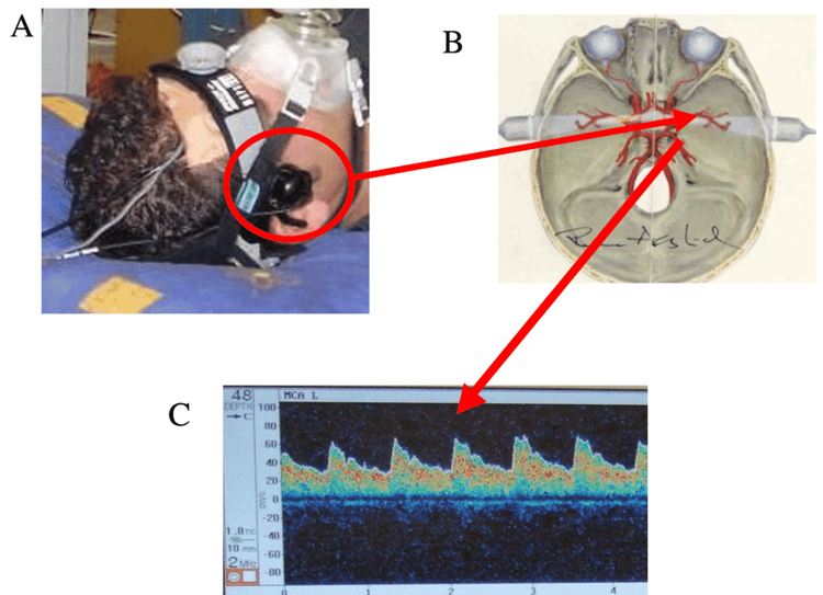
At this point, a transcranial doppler ultrasound can help evaluate degenerative conditions, blocked arteries, or other problems that affect blood flow to the brain. As a non-invasive imaging test, ultrasound uses a transducer that is moved around to different areas of the skull, where ultrasound waves bounce off red blood cells traveling through blood vessels. Accordingly, the speed of the blood flow will be measured without creating any images like other types of ultrasound. At the same time, the transcranial doppler ultrasound kit is also portable, so it can be performed at bed and the patient is fully awake during the procedure.

Siêu âm doppler xuyên sọ
2. Who should be ordered to perform transcranial doppler ultrasound?
Transcranial doppler ultrasound should be recommended for patients with the following risk factors:High blood cholesterol levels: People with elevated levels of triglycerides or cholesterol in the blood are more likely to have narrowing of the blood vessels. brain due to the accumulation of plaque on the walls of the arteries. Cardiovascular disease: A person diagnosed with cardiovascular disease may already have damaged arteries that will have a significant effect on the blood supply to the brain. Diabetes: This condition can lead to nerve damage and kidney disease, which can cause abnormally high blood pressure. At this time, high blood pressure will increase the risk of stroke.

Người có bệnh lý đột quỵ nhồi máu não
3. How is transcranial doppler ultrasound performed?
Before performing transcranial doppler ultrasound, the patient does not need any special preparation, including diet or prior fasting.During the procedure, the patient remains fully awake and lying in bed. A small device called a transducer, which is connected to a laptop computer, provides the sonographer or doctor with data about blood flow in the blood vessels of the brain. It is possible that one or more transducers will be placed directly on the patient's skin with a small amount of gel to facilitate the ultrasound. The technician will apply gel and place the transducer on the temple, the base of the skull at the back of the neck, or over the patient's closed eyelids.
At each location on the skull, the technician will adjust the orientation of the transducer to direct the ultrasound waves toward the blood vessels being examined. A single transcranial doppler ultrasound can take anywhere from 30 minutes to 1 hour to complete. After that, the gel on the skin can be easily washed off and the patient can sit up and return to normal activities.

Mô phỏng quá trình siêu âm doppler xuyên sọ
4. What are the results of transcranial doppler ultrasound?
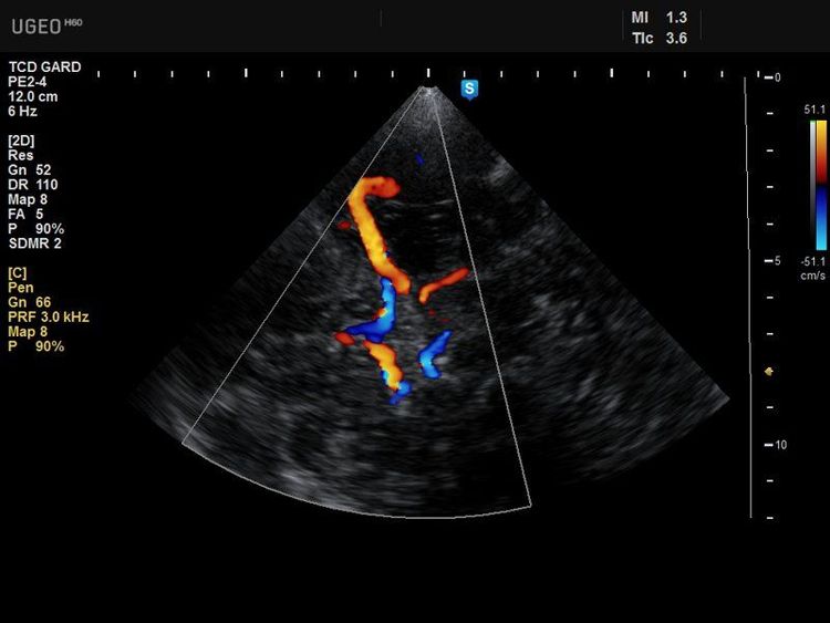
A result of a normal transcranial doppler ultrasound is when blood flow to the brain is normal, with no narrowing or blockages in the blood vessels to the brain and in the brain.Conversely, an abnormal transcranial doppler result is when anything is narrowing or clogging the arteries in the brain. This condition is seen in the following conditions:
Stroke or transient ischemic attack Subarachnoid hemorrhage Brain aneurysm Increased intracranial pressure Sickle cell anemia

Kết quả của siêu âm doppler xuyên sọ
In summary, thanks to its advantages and benefits, transcranial doppler ultrasound is a useful tool to help survey the cerebrovascular system. Thereby, patients are better treated and screened, contributing to the protection of brain parenchyma health in particular and comprehensive health in general.
With more than 14 years of working in the field of BSCK II imaging, Khong Tien Dat is currently a radiologist at Vinmec Ha Long International Hospital.
To register for examination and treatment at Vinmec International General Hospital, you can contact Vinmec Health System nationwide, or register online HERE
Recommended video:
Health check recurring at Vinmec: Protect yourself before it's too late!
LEARN MORE
Evaluation of pulmonary artery pressure by Doppler echocardiography The role of uterine Doppler ultrasound Suture and ablation of hemorrhoids under Doppler ultrasound guidance


