This is an automatically translated article.
The article was written by Master, Doctor Mai Vien Phuong - Gastroenterologist - Department of Medical Examination & Internal Medicine - Vinmec Central Park International General Hospital. Doctor has nearly 10 years of experience in the field of Gastrointestinal Endoscopy.Optical staining endoscopic techniques in particular and advanced endoscopy in general enable endoscopists to detect and characterize precancerous and neoplastic lesions and predict mucosal inflammation accurately. than conventional white light endoscopy.
1. Overview
Inflammatory bowel disease (IBD) includes 2 main types, ulcerative colitis (UC) and Crohn's disease (CD). It is now accepted that both groups of patients are at increased risk of developing cancer-associated colitis (CAC). Chronic inflammation of the gastrointestinal mucosa is an important factor for the development of inflammatory bowel disease-associated cancers (CACs). Therefore, chronic use of anti-inflammatory drugs has been suggested to reduce the risk of colitis.However, most studies cannot prove that anti-inflammatory drugs commonly used to treat inflammatory bowel disease have chemopreventive effects against cancer. Therefore, new efforts have been made to better define the development of mucositis in IBD in order to establish more effective targeted and preventive therapies.
Recently, Günther et al. have shown that the cysteine protease caspase 8 is critically involved in regulating intestinal homeostasis and protecting intestinal epithelial cells from TNF-induced necrotic cell death. -α-induced, thus suggesting new treatments in inflammatory bowel disease. It is clear that new and more advanced endoscopic imaging techniques are needed to monitor inflammatory bowel disease. In recent years, emerging endoscopic imaging techniques have been introduced, allowing more detailed analysis of mucosa and submucosa. This article describes the concept of advanced endoscopic imaging for the diagnosis and characterization of Crohn's disease.

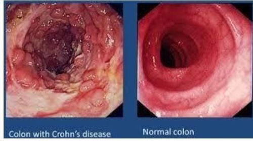
Hình ảnh nội soi của bệnh Crohn
2. Advanced endoscopic imaging methods
2.1. Magnifying endoscopy and dye-based staining endoscopy Magnifying endoscopy uses a movable lens to vary the magnification level up to 150x, thus allowing mucosal surface characterization (eg. For example, the surface pattern classification of colon polyps was proposed by Kudo et al. 1996).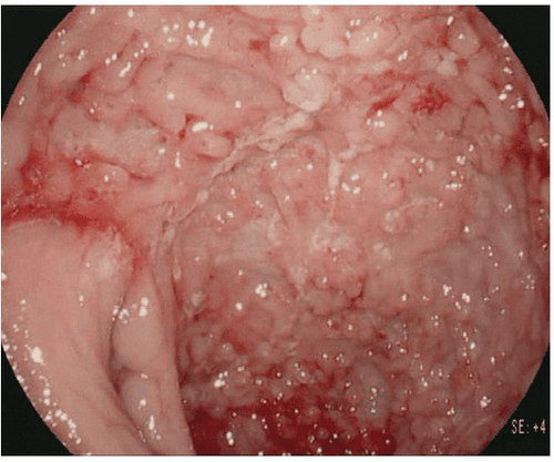
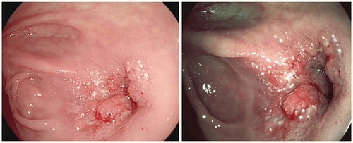
Nội soi ánh sáng trắng độ nét cao của bệnh Crohn nặng với nhiều vết loét dọc và xuất huyết tự phát của niêm mạc bị viêm.
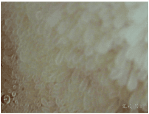
Nội soi độ phóng đại cao sử dụng i-scan với sự cải thiện bề mặt và nâng cao tông màu cho thấy rõ từng nhung mao ruột có hình dạng và kích thước bình thường ở một bệnh nhân mắc bệnh Crohn chưa ổn định.
2.2. Dye-free endoscopy (optical staining) Dye-free endoscopy is divided into optical pigmented endoscopy including narrow band imaging (NBI; Olympus, Tokyo, Japan) and endoscopy. Virtual pigments include i-scan (Pentax, Tokyo, Japan) and Fujinon intelligent color enhancement (FICE; Fujinon, Tokyo, Japan). While NBI is based on optical filters inside the endoscope light source, narrowing the spectral transmission bandwidth, thereby enhancing vascularity, i-scan and FICE use digital post-processing to estimate the spectrum. calculated to produce better tissue contrast.

Vết loét lớn được kiểm tra bằng hình ảnh dải hẹp. NBI hình dung kiến trúc bề mặt không đều và cấu trúc mạch máu bị xáo trộn. Mô bệnh học khẳng định chẩn đoán ung thư.
Hỉnh ảnh i-scan: (a) làm nổi bật hình ảnh nội soi ánh sáng trắng có độ nét cao về hẹp miệng nối ở một bệnh nhân bị bệnh Crohn. (b) hiển thị nội soi sắc tố ảo với i-scan để mô tả rõ hơn tính chất nghiêm trọng của vị trí hẹp. Hình ảnh i-scan cho thấy các tổn thương mạch máu và xơ tăng cường kèm theo tình trạng viêm đang hoạt động.
Recently, Vden Broek and colleagues performed a randomized crossover trial in which UC patients underwent both inflammatory bowel disease white light colonoscopy and high definition (HD) in randomized order. course. It was shown that NBI did not improve cancer detection in patients with ulcerative colitis compared with HD colonoscopy. In addition, the NBI proved unsatisfactory in distinguishing cancerous from noncancerous mucosa. The same group evaluated the value of three-way endoscopic imaging for surveillance in ulcerative colitis. 50 patients underwent surveillance colonoscopy and each colon segment was examined twice, once with automated fluorescence imaging (AFI) and once with white light endoscopy, in random order . All detectable lesions were examined by NBI for Kudo surface pattern analysis and additional randomized biopsies were performed. AFI improves cancer detection and reduces random biopsy yield. Analysis of pit samples by NBI was moderately accurate for histological prediction, while AFI coloration was valuable in excluding the presence of tumors.
3. The role of NBI optical staining endoscopy in the assessment of dysplastic inflammatory lesions in the colon
Kudo et al analyzed mucosal vascularization (MVP) patterns in ulcerative colitis using both conventional colonoscopy and NBI. In this study, NBI colonoscopy was more accurate for determining the degree of inflammation in patients with unstable ulcerative colitis than the histological gold standard.In another study, the same group performed a pilot study of magnified colonoscopy with NBI for the diagnosis of dysplasia in ulcerative colitis. The surface pattern is defined as honeycomb-like, villi-like, or meander-like. By taking into account the surface model, the rate of positive dysplasia was higher in the meander model than in the honeycomb or villi model group.
Therefore, the tortuous pattern identified by NBI colonoscopy may be a clue to identify dysplasia during surveillance for ulcerative colitis. Recently, it has also been shown that NBI appears to be a less time-consuming and equally effective alternative to chromoscopy for detecting intraepithelial neoplasia. NBI resulted in a significantly lower rate of false-positive biopsies and a similar true-positive rate. However, given the incidence of NBI lesions and patient omission rates, NBI cannot be recommended as the standard surveillance technique in inflammatory bowel disease. Another pilot study addressed the question of whether NBI could successfully assess mucosal angiogenesis in inflammatory bowel disease. In endoscopically normal but NBI-positive areas, there was a significant increase in mucosal angiogenesis. Thus, NBI may enable in vivo imaging of intestinal neoplasia in patients with inflammatory bowel disease.
4. The role of high-definition (HD) endoscopy in the diagnosis of Crohn's disease
There are only limited data available on the phantom chromoscopic technique in inflammatory bowel disease. To date, only one study has evaluated FICE in the inflammatory bowel disease setting, showing that the system cannot improve the detection or identification of ulcers and erosions in CD. Another study compared methylene blue appendectomy (0.1%) and HD colonoscopy using i-scan. HD colonoscopy with and without i-scan is feasible to detect many small lesions but pigmentoscopy can even increase the number. Furthermore, i-scan can predict cancer as accurately as chromoscopy. A randomized controlled study evaluated HD colonoscopy in combination with i-scan or standard video endoscopy for the detection of colorectal lesions. HD colonoscopy with i-scan detected significantly more colorectal cancer patients (38%) than standard resolution colonoscopy (13%). Significantly more cancerous lesions and more flat adenomas can also be detected by HD endoscopy with i-scan. Recently, our group successfully investigated the effect of i-scan in predicting mucositis in IBD. The patient underwent a total colonoscopy and was examined with both HD (Group A) and HD plus i-scan colonoscopy (Group B). The agreement between the endoscopic prediction of disease severity and histological outcome was 65% in group A and 88% in group B. When endoscopic predictions of inflammatory activity were compared with tissue results From the respective samples, we found agreement in 42% in group A and 85% in group B. Compared with histological results of inflammation, i-scan allowed a significantly more accurate diagnosis. mucosal inflammation compared with conventional colonoscopy.Dye-free chromoscopy has the potential to replace conventional dye-based chromoscopy for lesion detection and disease severity assessment in inflammatory bowel disease. Virtual pigmentary endoscopy with i-scan is superior to standard video colonoscopy for colorectal cancer detection. I-scan significantly improved the diagnosis of the severity and extent of mucositis in IBD patients.

Nội soi HD với i-scan phát hiện nhiều bệnh nhân ung thư đại trực tràng hơn đáng kể
5. Conclusion
State-of-the-art endoscopic imaging in inflammatory bowel disease has seen a major innovation revolution in the past 10 years, including both live and virtual chromatoscopic endoscopy. By using capsule endoscopy or balloon-assisted small bowel endoscopy, endoscopists can now assess the entire small intestine and perform endoscopic therapies in a place that has never been visited before. via . Furthermore, recent advances in endoscopic imaging now allow endoscopists to obtain real-time in vivo histology during ongoing endoscopy using microscopy or cytoskeletal endoscopy. . These emerging imaging modalities allow the endoscope to detect and characterize precancerous and neoplastic lesions and predict mucosal inflammation more accurately than conventional white light endoscopy often. Recent efforts have been made to introduce molecular imaging in inflammatory bowel disease by highlighting inflammatory cells and specific receptors. The results of these trials are highly anticipated and may open new avenues for diagnostic and therapeutic strategies in inflammatory bowel disease.Vinmec International General Hospital is a prestigious address trusted by many patients in performing diagnostic techniques for digestive diseases, diseases that cause chronic diarrhea, Crohn's disease... Along with that, at Vinmec Hospital, screening for gastric cancer and gastric polyps is done through gastroscopy with Olympus CV 190 endoscope, with NBI (Narrow Banding Imaging) function for The imaging results of mucosal pathology analysis are clearer than conventional endoscopy, detecting ulcerative colitis lesions, early gastrointestinal cancer lesions... Vinmec Hospital with facilities Quality and modern equipment and a team of experienced experts, always dedicated to medical examination and treatment, customers can rest assured with gastroscopy and esophagogastroduodenoscopy at Vinmec International General Hospital. .
Please dial HOTLINE for more information or register for an appointment HERE. Download MyVinmec app to make appointments faster and to manage your bookings easily.
References:1. Ullman TA, Itzkowitz SH. Intestinal inflammation and cancer. Gastroenterology. 2011;140(6):1807–1816. [PubMed] [Google Scholar]
2. Farraye FA, Odze RD, Eaden J, Itzkowitz SH. AGA technical review on the diagnosis and management of colorectal neoplasia in inflammatory bowel disease. Gastroenterology. 2010;138(2):746–e4. [PubMed] [Google Scholar]
3. Günther C, Martini E, Wittkopf N, et al. Caspase-8 regulates TNF-alpha , epithelial necroptosis and terminal ileitis. Nature. 2011;477(7364):335–339. [PMC free article] [PubMed] [Google Scholar]
4. Neumann H, Vieth M, Langner C, Neurath MF, Mudter J. Cancer risk in IBD: how to diagnose and how to manage DALM and ALM. World Journal of Gastroenterology. 2011;17(27):3184–3191. [PMC free article] [PubMed] [Google Scholar]
5. Rutter MD, Saunders BP, Wilkinson KH, et al. Thirty-year analysis of a colonoscopic surveillance program for neoplasia in ulcerative colitis. Gastroenterology. 2006;130(4):1030–1038. [PubMed] [Google Scholar]
6. Helmut Neumann, Klaus Mönkemüller. Advanced Endoscopic Imaging for Diagnosis of Crohn's Disease, Gastroenterol Res Pract. 2012; 2012: 301541