This is an automatically translated article.
This article was professionally consulted by a Doctor of Radiology, Department of Radiology - Vinmec Central Park International General Hospital.The left ventricle is one of the four chambers of the heart, the left ventricle is responsible for the extremely important job of pumping blood into the circulatory system to feed the whole body. Thus, once the left ventricular systolic function declines, it will cause anemia to nourish the body, thereby causing pathological conditions in other organs.
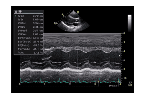
Therefore, the assessment of left ventricular systolic function is very important. Today, doctors mainly rely on Doppler echocardiography to evaluate heart function in general and left ventricular function in particular.
1. Location and function of the left ventricle
Our heart consists of four chambers which are:Right atrium: is the upper, right chamber of the heart. Left atrium: the upper left chamber of the heart. Right ventricle: the lower right chamber of the heart. Left ventricle: The lower left chamber of the heart. The left ventricle has the thickest layer of myocardium in the chambers of the heart. The circulation in our body is as follows: blood after receiving oxygen in the lungs will follow the pulmonary veins to the left atrium. From the left atrium, blood flows down the left ventricle when the mitral valve is open during diastole (when the heart relaxes). During systole (when the heart contracts) blood from the left ventricle will be pumped into the aorta, from which blood will travel throughout the body to nourish the organs.
After metabolism, blood will bring CO2 and metabolic products of cells through the venous route to the right atrium. From the right atrium, blood flows to the right ventricle when the tricuspid valve is open during diastole. During systole, blood from the right ventricle will be pumped into the pulmonary artery, to the lungs to excrete CO2 and receive O2. Blood continues to return to the left atrium, continuing the cycle without stopping.
Thus, the left ventricle takes on a very important role, which is to pump blood containing O2 to the organs in the body. Therefore, the assessment of left ventricular systolic function is extremely important.

Tâm thất trái đảm nhiệm vai trò là bơm máu có chứa O2 đến các cơ quan trong cơ thể
2. The role of Doppler echocardiography in diagnosing left ventricular systolic function
Doppler ultrasound is a type of imaging probe that helps to examine moving objects with a transducer that emits and receives ultrasound waves. Doppler echocardiography can measure blood flow velocity readings at different locations in the heart chambers. The advantage of echocardiography is that it can be performed in the ultrasound room, or at the bedside in case of an emergency. However, it also has a disadvantage that in obese patients, the chest wall is thick, the image quality is limited.Cardiac doppler echocardiography will provide us with indicators to evaluate left ventricular systolic function, which are:
Cardiac output CO Index dp/dt left ventricular ejection time (PET) Ejection time ( ET) Systolic S wave 2.1. Cardiac Output CO Cardiac output CO is the amount of blood pumped by the left ventricle into the aorta in 1 minute.
Calculated according to the following formula:
CO = SV x Heart rate .
SV (Stroke Volume) is the amount of blood pumped into the aorta during each heartbeat, also known as stroke volume. Left ventricular systolic function is normal when CO is between 4-6 l/min.
2.2. Left ventricular dp/dt index Left ventricular dp/dt index represents the process of increasing left ventricular pressure over time. This index can only be calculated when mitral regurgitation is present. Diagnosis of left ventricular systolic function and LV dp/dt index is as follows:
dp/dt > 1200: normal left ventricular systolic function. 800 < dp/dt < 1200: left ventricular systolic function is moderately reduced. dp/dt < 800: left ventricular systolic function is severely reduced. 2.3. Pre-ejection time (PET) The pre-ejection time measured on cardiac doppler echocardiography was calculated from the base of the R wave to when the aortic valve opened.
The longer the ejection time, the lower the left ventricular systolic function.
Preload time is affected by:
Heart rate Preload 2.4. Ejection time (ET) Ejection time is measured as the time interval between two open and close clicks of the aortic valve.
The shorter the ejection time, the lower the left ventricular systolic function.
PET/ET > 0.65 is equivalent to EF < 30%.
Ejection time is affected by:
Heart rate Preload Afterload 2.5. S-systolic wave S-systolic wave, V(S) reflects the contractile function of the left ventricle.
V(S) > 8 cm/s is a normal left ventricular contractile function.
V(S) < 8 cm/s means reduced left ventricular systolic function, coronary artery disease, old age.
Doppler echocardiography provides doctors with cardiac output parameters CO, left ventricular dp/dt index, ejection time (PET), ejection time (ET), systolic S wave. Through these indicators, the doctor can assess whether the patient's left ventricular systolic function is normal or not.

Thông qua siêu âm doppler tim bác sĩ sẽ có thể đánh giá chức năng tâm thu thất của bệnh nhân có bình thường hay không
If you have a need for consultation and examination at the Hospitals of the National Health System, please book an appointment on the website for service.
Please dial HOTLINE for more information or register for an appointment HERE. Download MyVinmec app to make appointments faster and to manage your bookings easily.