This is an automatically translated article.
The article was professionally consulted by Doctor Head of Department of Medical Examination & Internal Medicine, Vinmec Hai Phong International General Hospital
Cardiomyopathy is a major cause of heart failure, with a poor prognosis and high mortality rate. Early diagnosis is important in the detection and treatment of the disease. In which, computed tomography is a technique for diagnosing cardiomyopathy with high accuracy, being widely applied today.
1. General understanding of cardiomyopathy
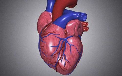
1.1 What is cardiomyopathy? Cardiomyopathy is a term that describes an abnormal heart muscle condition, which is a progressive heart disease that causes the heart to expand, thicken, or stiffen abnormally. In patients with cardiomyopathy, it is difficult for the heart to pump and supply blood to the rest of the body. This disease has a poor prognosis, the mortality rate after 5 years is 35% and after 10 years is 70%.
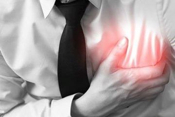
1.2 Symptoms of cardiomyopathy In the early stages, people with cardiomyopathy often have no symptoms. However, as the disease progresses, symptoms of the disease will appear. These are:
Shortness of breath on exertion or even at rest; Leg swelling: ankles and feet; Cough while lying down; Abdominal distention for fluid retention; Tired; Abnormal heartbeat: fast, pounding, or fluttering; Palpitations, chest pain; Dizziness, fainting; High Blood Pressure. A stroke if a blood clot forms in the left ventricle, the skin dilates and the clot breaks, disrupting blood flow to the brain. 1.3 Causes of cardiomyopathy There is no exact cause of cardiomyopathy. Some factors that increase your risk include:
Family history of cardiomyopathy, heart failure or sudden cardiac arrest; Persistent high blood pressure; Have chronic heart palpitations ; Damage to heart tissue from a previous myocardial infarction; Have heart valve disease ; Having metabolic disorders such as diabetes, obesity, thyroid disease; Complications of pregnancy (perinatal cardiomyopathy); Deficiency of essential vitamins and minerals (eg thiamin - vitamin B1); Drinking alcohol for a long time; Use of amphetamines, cocaine, or anabolic steroids; Using certain chemotherapy drugs and radiation for cancer; Accumulation of iron in the heart muscle (iron overload); Having certain infections can damage the heart and trigger cardiomyopathy; Inflammation that causes clumps of cells to grow in the heart and other organs (sarcoidosis); Have a disorder that causes abnormal protein buildup (amyloidosis); Connective tissue disorders.
1.4 Diagnostic method of cardiomyopathy The doctor will diagnose cardiomyopathy through methods such as:
Chest X-ray: showing images of the heart to help assess whether the heart muscle is dilated; Echocardiography: A method that uses sound waves to create images of the heart. From these images, the doctor can check the size, function of the heart and movement when the heart beats; Electrocardiogram (ECG); Check your heart rate, blood pressure, and breathing rate during strenuous walking on the treadmill. Cardiac catheterization; Computed tomography (CT); Magnetic resonance imaging (MRI) ; Blood tests; Genetic testing or screening. 1.5 Treatment of cardiomyopathy The choice of treatment for cardiomyopathy depends mainly on the extent of the damage to the heart and the external symptoms. The goal of treatment is to help the heart work more efficiently and prevent further damage. Cardiomyopathy is difficult to cure completely but can be controlled by:
Change to a healthier lifestyle: avoid alcohol and cocaine use; control high blood pressure, high cholesterol and diabetes; Healthy eating; increase exercise and sports; reduce stress; get enough sleep; quit smoking,...; Use medications to treat high blood pressure, prevent fluid retention, keep your heart rate normal, prevent blood clots, and reduce inflammation; Using pacemakers and defibrillators; Heart transplant surgery is the last resort if the above methods do not work.

2. Computed tomography - an accurate method of diagnosing cardiomyopathy
Computerized tomography (CT scan) is an imaging method that uses X-rays to be scanned horizontally through a part of the human body.
Accordingly, in cardiac tomography, the localized location of the X-ray is the heart. By taking advantage of the measurements of X-rays, computed tomography (CT) scans will produce multiple imaging results from different angles on the heart, helping doctors make an accurate diagnosis of the patient's condition.
This is the most accurate and fast diagnostic technique for cardiovascular diseases through detailed images of the heart and arteries. In addition, based on the extent and location of plaque buildup in the coronary arteries, doctors also easily determine the patient's risk of heart disease. In particular, tomography easily diagnoses some cardiomyopathies such as hypertrophic cardiomyopathy, thereby helping doctors soon give the necessary treatment indications for patients.
3. Why should CT scan to diagnose cardiomyopathy at Vinmec?
Currently, Vinmec Hai Phong International General Hospital is applying computed tomography of coronary arteries and hearts without beta block for patients with cardiomyopathy and other cardiovascular diseases.
Choosing computerized tomography techniques at Vinmec Hai Phong helps customers get the earliest and most accurate diagnosis thanks to:
Modern machinery system, meeting international standards: CT scanner 640 slices Aquilion One ( Vision Edition) fast shooting speed, possessing the latest AIDR 3D ray dose reduction technology, allowing 75% reduction of the dose to the patient; The machine is capable of synthesizing 320/640 slices in one revolution at a speed of 0.275*s/0.35s; The system uses a 160mm wide acquisition Detector with 320 rows of Detector allowing to capture the heart, brain,... in one rotation, shortening the diagnostic time; iStation helps to display ECG signals, guide breathing on Gantry; SureExposure 3D continuously adjusts the imaging current during the helical scan along the three spatial axes of X, Y and Z according to the patient's body shape, reducing radiation dose to the lowest possible level. Voice guidance to the patient speak and system (Voice link); Boost3DTM allows dose reduction for areas with high X-ray absorptiveness such as the shoulder area for high-precision image acquisition; 3D color image processing (surface reconstruction, volume reconstruction, MIP, MPR, curved MPR, Cine) Vinmec Hai Phong's team of highly qualified and experienced doctors, namely: Master, Doctor Doan Xuan Sinh: has 17 years of experience in diagnostic imaging, especially in the field of pediatric imaging; Specialist Doctor I Nguyen Dinh Hung: has more than 10 years of experience in the field of diagnostic imaging (ultrasound, CT and MRI); Doctor, Doctor Pham Quoc Thanh: has 13 years of experience in the field of diagnostic imaging, has received extensive training and participated in many national and international scientific conferences on diagnostic imaging. Cardiomyopathy can be life-threatening if not detected and treated early. Therefore, when there are warning symptoms of cardiomyopathy, patients should go to Vinmec Hai Phong Hospital to have the best experience of coronary artery and cardiac computed tomography without beta block medication.
Please dial HOTLINE for more information or register for an appointment HERE. Download MyVinmec app to make appointments faster and to manage your bookings easily.


