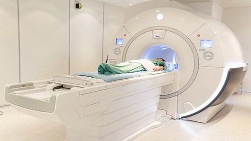
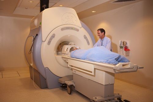
This is an automatically translated article.
The article is expertly consulted by Master, Doctor Hoang Van Lan Duc - Doctor of Radiology - Department of Diagnostic Imaging and Nuclear Medicine - Vinmec Times City International General Hospital.To diagnose diseases in the cervical spine, many patients are prescribed magnetic resonance imaging of the cervical spine without injection of magnetic contrast. This is a diagnostic method with a very high accuracy and safety rate, so it is widely applied today.
1. Benefits of having an MRI of the cervical spine
Magnetic resonance imaging is an imaging technique that does not use X-rays, is non-invasive, and does not contain ionizing radiation. It is the most effective method when compared with other imaging methods because of its ability to clearly and detailed evaluation of the cervical spine. In particular, this method can also help doctors assess the condition of the spinal cord and nerves (something other diagnostic techniques cannot do).Magnetic resonance imaging can evaluate areas that are hidden by bone or difficult to see in other imaging methods. This is also a particularly useful technique in diagnosing compression, acute myelitis (clinical manifestations of paralysis or numbness). Spinal diseases such as damage to discs, vertebral bodies, spinal cord and soft tissues around the spine,... can all be diagnosed accurately by MRI.
2. Indications for magnetic resonance imaging of the cervical spine without injection of magnetic contrast agents
In case the patient presents with severe arm pain, unresponsive to long-term treatments; In case the patient has long-term neck pain, shoulder pain, poor response to treatment;
Người bệnh bị đau cổ vai gáy và đáp ứng điều trị kém cần được chụp cộng hưởng từ cột sống cổ
3. Contraindications to magnetic resonance imaging of the cervical spine without injection of magnetic contrast agents
3.1 Absolute contraindications Severe patients, requiring resuscitation equipment on the body; People who have been implanted with electronic devices on the body such as cochlear implants, automatic drug pumping devices under the skin, pacemakers, anti-vibration machines,...; People with metal clips in the blood vessels, intracranial or orbit for less than 6 months; Pregnant women under 12 weeks of age. Pregnant women over 12 weeks can have an MRI without affecting the fetus.
Phụ nữ mang thai dưới 12 tuần tuổi, tuyệt đối không chụp cộng hưởng từ cột sống cổ
4. Perform magnetic resonance imaging of the cervical spine without injecting magnetic contrast
4.1 Preparation of Implementation Personnel: Including specialists, nursing assistants and imaging technicians; Technical facilities: Including MRI machine; film, film printers and image storage systems; Patient: Received detailed explanation of necessary technical information by the doctor; checked for contraindications; no need to fast; change the clothes of the magnetic resonance imaging room, remove the items on the list of contraindications; bring the doctor's request for magnetic resonance imaging or medical records if available; 4.2 Conducting MRI of cervical spine Patient position: Lying supine on the scanning table; Doctor's choice - position the receiver coil. Then, the doctor moves the table into the correct magnetic field of the magnetic resonance imaging machine and locates the imaging area; Location capture; Capture the T1W, T2W vertical and transverse T2W pulse sequences through the required position. Stir can be shot vertically if needed; At the end of the magnetic resonance imaging process, the medical staff removed the signal receiver coil and invited the patient out of the imaging room, waiting for the results; The technician will process the images, print the film and transfer the obtained images and data to the doctor; The doctor performs image analysis and makes a diagnosis.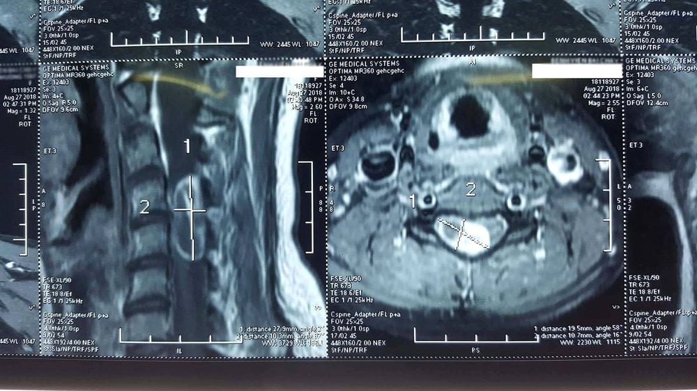
Hình ảnh chụp Cộng hưởng từ (MRI) đột sống cổ trên bệnh nhân có khối u tủy cổ
Vinmec International General Hospital is a high-quality medical facility in Vietnam with a team of highly qualified medical professionals, well-trained, domestic and foreign, and experienced.
A system of modern and advanced medical equipment, possessing many of the best machines in the world, helping to detect many difficult and dangerous diseases in a short time, supporting the diagnosis and treatment of doctors the most effective. The hospital space is designed according to 5-star hotel standards, giving patients comfort, friendliness and peace of mind.
Master. Doctor Hoang Van Lan Duc was formally trained and graduated with a Master's degree from Hanoi Medical University. The doctor has more than 10 years of experience in the field of imaging, especially in diagnosing with emergency internal, surgical, abdominal, thoracic, musculoskeletal, nerve and thyroid, breast, ...
To register for an examination at Vinmec International General Hospital, you can contact the nationwide Vinmec Health System Hotline, or register online HERE.


