This is an automatically translated article.
Currently, laparoscopic right extension colectomy combined with lymph node dissection is being used for patients with tumors or colon cancer, this method helps patients have many advantages over other methods. other law.
1. Learn about laparoscopic right colonectomy with lymph node dissection
Laparoscopic extended colectomy is the removal of 10 to 15 cm of the ileum, cecum, ascending colon, right transverse colon, and corresponding mesentery performed by laparoscopic technique. Restoration of the gastrointestinal tract by anastomosis of the ileum to the colon through a small opening of the abdominal wall.
Lymph node dissection is the resection of a right colonic tumor as determined by resection of the ileocecal vessel of the colic, right colon, middle colon, and proximal right gastric omental artery.
2. Indications and contraindications
Indications for right dilated colectomy are common for cancer of the hepatic flexure colon or in the transverse colon near the hepatic flexure. Contraindicated for patients with severe coagulopathy, cardiopulmonary and respiratory diseases; when the tumor is too large to invade widely; Patients who are too fat and weak to bear, should consider when having colectomy surgery.
3. What to prepare before surgery?

Trước khi mổ cần nhịn ăn
Colon needs to be cleaned of stool before surgery. The patient only drank milk on the first day and sugar water on the second day. Cleanse the intestines by means of enemas or enemas, depending on the instructions and instructions of the doctor. Absolutely fasting, bathing with sterile soap the night before surgery. Stop taking anti-pain, anti-inflammatory, anticoagulant drugs. Tell your doctor if you have not cleaned your bowels, or if you have any discomfort before surgery.
4. Procedure for performing surgery
Step 1: Investigate and evaluate liver and peritoneal metastases, then find the ileocolic bundle, create two windows above and below this vessel Step 2: Evaluate the ureter Step 3: Surgery along the vein superior mesenteric and from the base of the ileocecal artery of the colon. Clamping and excision of the ileo-colic vessels. The right colic artery must be cut, then the right colic vein is removed, and the right gastric omental vein is left close to the base of the vein of Henle. Step 4: The great omentum is released into the omentum harem. Resection of the great omentum is directed from the middle to the periphery and to the right gastric artery. Separation of the duodenum and head of the pancreas from the transverse mesentery. Step 5: Release the hepatic flexure from left to right or from right to left. Step 6: Cut the terminal ileum with a stapler and the cecum is moved. Step 7: Using the trocar on the right side and above the pubic bone, the transverse colon is raised to visualize the duodenum and the middle colonic vessels. Step 8: Resection of the great omentum continues to the left by lifting the great omentum with a pin and extending it completely into the omentum posterior, avoiding pulling the spleen. Release the splenic flexure by pulling the colon toward the midline and cutting the fixation, pulling the splenic flexure from the posterior wall of the peritoneum. Step 9: Left colon cross section with stapler, from right trocar hole. The two mesenteric margins of the ileum and descending colon were elevated and the lateral ileocolonic anastomosis was performed with a stapler inserted through the trocar hole in the pubic bone. Two perforated holes for stapler mounting are sewn together with double-layer monosilk thread in the body. The open mesentery can be left open. Step 10: Put the specimen in a dedicated bag, cover the incision through the opening of the trocar umbilicus or on the pubic bone. Close the skin with the thread 4-0 .
5 Monitoring and handling of accidents

Theo dõi,chăm sóc cho bệnh nhân và cho bệnh nhân uống thuốc kháng sinh sau khi mổ
Follow-up
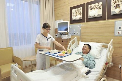
Care after surgery such as gastrointestinal surgery Rehydrate the patient, monitor the drain, and urine after surgery Give the patient antibiotics after surgery After surgery If the patient is able to pass the stool, then start feeding them as thin as porridge or milk. Treatment of complications
Large tumors should not be operated on laparoscopically, must be operated on. During surgery, bleeding may occur, find out the reason for duodenal perforation, cut the ureter, and find a specific treatment. After surgery, the patient shows signs of bleeding in the abdomen, if necessary, follow up, then re-operate, open or laparoscopically. Surgery to find the cause and check the residual abscess in the abdomen needs to be cleaned and drained. Vinmec International General Hospital with a system of modern facilities, medical equipment and a team of experts and doctors with many years of experience in neurological examination and treatment, patients can completely rest in peace. examination and treatment center at the Hospital.
To register for examination and treatment at Vinmec International General Hospital, you can contact Vinmec Health System nationwide, or register online HERE.
MORE
Advantages of gastroscopy and colonoscopy with anesthesia Laparoscopic right and left colectomy in the treatment of rectal cancer The need for colorectal cancer screening


