This is an automatically translated article.
The article was professionally consulted by Specialist Doctor I Nguyen Truong Duc - Radiographer - Department of Diagnostic Imaging and Nuclear Medicine - Vinmec Times City International General Hospital.CT scan of the lumbar spine with intravenous contrast is often indicated to evaluate diseases of the spine (such as tuberculosis, tumor, inflammation...) that have damage to the soft tissues around the spine or Evaluation of blood vessel damage in the lumbar spine after trauma.
Diseases in the lumbar spine such as herniated discs, spine spines, arthritis, spondylolisthesis,... can all be detected like a CT scan of the lumbar spine without contrast injection. After diagnosis results are available, patients can be detected and treated early and effectively.
1. Common diseases of the lumbar spine
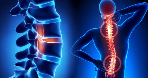
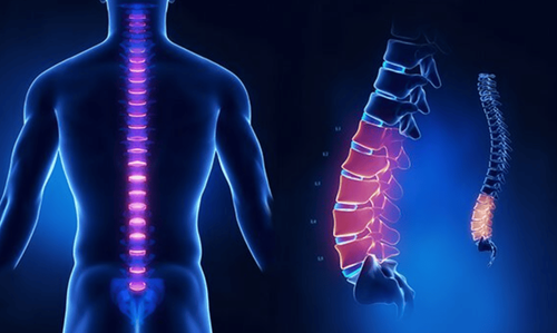
The lumbar spine is the central structure of the human body, supporting the weight of the entire body. According to a survey, up to 80% of the world's population has at least once suffered from diseases related to the lumbar spine.Some common diseases in the lumbar spine, causing pain symptoms include:
Degeneration of the lumbar spine due to aging or having been injured in an accident, specific occupation, playing sports, .. .;
Herniated disc ;
Tuberculosis of the spine ;
Spondylitis ;
Cancer.
If the pain in the lumbar spine is mild, it will affect the patient's daily activities, making it difficult to stand up, sit down, turn,... If the pain is caused by a herniated disc, it can be causing sciatica pain, over time leading to muscle atrophy of thighs and legs, urinary disorders, even leaving severe sequelae such as paralysis.
To detect diseases in the lumbar spine, the doctor may appoint the patient to perform a CT scan of the lumbar spine with contrast injection.

Chụp CLVT có tiêm thuốc cản quang giúp phát hiện sớm bệnh thoái hóa cột sống thắt lưng
2. The procedure for taking a CT scan of the lumbar spine with contrast injection
This is a technique that uses a computerized tomography machine to create images of the lumbar spine, helping to evaluate the damage of bones, discs as well as the spinal canal and adjacent components. When combined with iodinated contrast injection, intravenous CT scan of the lumbar spine helps to evaluate inflammatory diseases, tuberculosis, spinal tumors or vascular lesions,...2.1 Indications and contraindications
● Indications: Suspected cases of trauma, inflammation, bone and soft tissue tumors of the lumbar spine; Cases of suspected vascular injury in the lumbar spine such as aneurysms, arteriovenous malformations...● Contraindications: This technique has no absolute contraindications. However, CT scanning of the lumbar spine is relatively contraindicated in pregnant women, patients with renal failure, or those who are allergic to iodinated contrast.
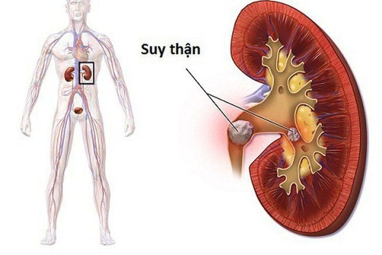
Bệnh nhân suy thận chống chỉ định chụp CLVT cột sống thắt lưng
2.2 Preparation before implementation
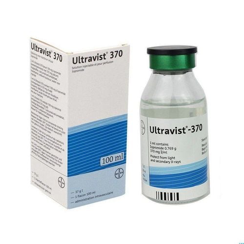
● Performers: Specialist doctors, nurses and radiology technicians;● Medical supplies: Needles, syringes of all sizes, syringes for electric pumps, trays, surgical forceps, physiological saline or distilled water, water-soluble iodine contrast agents, skin antiseptic solutions, gloves, hats, masks, cotton, gauze, medicine boxes and emergency equipment for contrast drug accidents;
● Technical facilities: Specialized electric pump, computer tomography machine, film, film printer and image storage system;
● Patient: Get a clear explanation of the procedure; need to fast and drink before 4 hours, can drink less than 50ml of water; remove necklaces, earrings and other metal objects; you may be given a sedative if you are too excited to stay still;
● Test sheet: Order of computed tomography scan and medical records, other imaging results, if any.
2.3 Technical implementation
● Patient position: The patient needs to lie on his back in the machine frame, with the shoulders as low as possible and the arms raised up along the body axis. Patient holds breath, does not swallow during CT scan;● Localization of the entire lumbar spine in 2 planes;
● Take the lateral positioning image, starting from the upper shore D12 to the lower shore S1;
● Set the scan program according to clinical requirements. The doctor can use cutting layers in the direction of the discs to evaluate herniated disc pathology or take a picture of the entire lumbar spine and use image processing software after taking it;
● Select film images on bone windows and disc windows;
● Take again after intravenous contrast injection.
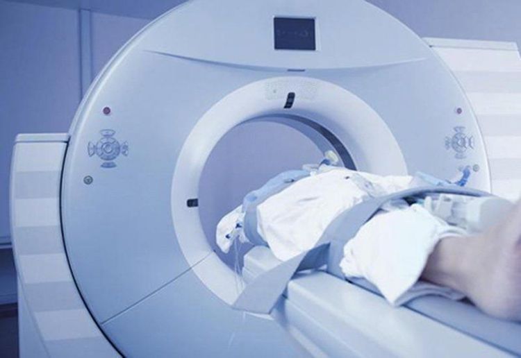
Hình ảnh bệnh nhân chụp cắt lớp vi tính cột sống thắt lưng
2.4 Evaluation of results
● Compare images before and after contrast injection, identify comorbidities; Background removal techniques can be used to reconstruct vascular images.● Evaluation of congenital abnormalities of the spine;
● Evaluate posterior arch injury, vertebral body injury (collapsed vertebrae, vertebral body rupture, vertebral body slip), damage to posterior vertebral body wall, traumatic hematoma, signs of disc herniation, partial injury soft groove,...;
● Evaluation of damage in degenerative spondylosis such as ligament degeneration, lateral joint block degeneration, spinal stenosis, spondylolisthesis due to degeneration,...
● Assess the degree of contrast enhancement of soft tissue lesions around the spine.
2.5 Risk of complications and treatment measures
● The technique may have to be repeated if there are some errors such as the patient not keeping still during the CT scan, not revealing the image clearly,...;● Contrast-related adverse events: These include mild reactions, moderate reactions, and severe anaphylaxis. Depending on each reaction, there will be appropriate treatment options according to the standard treatment protocol.
CT scan of the lumbar spine may have to be repeated if the patient does not follow the doctor's instructions during the technique. Therefore, in order for the CT scan process to take place smoothly and without taking much time, the patient should follow all instructions of the doctor.

Sốc phản vệ là một tai biến có thể xảy ra khi chụp CLVT cột sống thắt lưng
A system of modern and advanced medical equipment, possessing many of the best machines in the world, helping to detect many difficult and dangerous diseases in a short time, supporting the diagnosis and treatment of doctors the most effective. The hospital space is designed according to 5-star hotel standards, giving patients comfort, friendliness and peace of mind.
Doctor Duc has nearly 17 years of experience in diagnostic imaging at Hanoi E hospital. Currently, the doctor is working at the Department of Diagnostic Imaging - Vinmec Times City International Hospital.
To register for an examination at Vinmec International General Hospital, you can contact the nationwide Vinmec Health System Hotline, or register online HERE.


