This is an automatically translated article.
Posted by CKII Doctor Khong Tien Dat - Diagnostic Imaging Department - Vinmec Ha Long International Hospital
Magnetic resonance imaging of the lumbar spine has high contrast, good anatomical details, allowing accurate detection of morphological and structural lesions of the parts of the body being examined.
1. What is magnetic resonance imaging?
Magnetic Resonance Imaging (MRI) is an imaging technique considered as one of the revolutionary inventions in medical technology, using magnetic fields and radio waves. Magnetic resonance imaging (MRI) with high contrast, good anatomical detail allows accurate detection of morphological and structural lesions of the parts of the body being examined. The ability to reproduce good 3D images, without side effects, is increasingly widely specified for many different specialized applications.
2. When is magnetic resonance imaging of the lumbar spine?
Patients may be assigned to take MRI of the lumbar spine when:
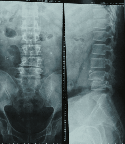
Back pain persists for unknown reasons or after carrying heavy objects; Numbness and tingling in extremities Have a history of cancer Loss of bladder or bowel control Back injury Inflammation, skin abscesses in the back are suspected to have spread to the lumbar spine.

Người bệnh đau lưng kéo dài
3. Purpose of lumbar spine magnetic resonance imaging
Magnetic resonance imaging of the lumbar spine can reveal the following groups of pathologies:
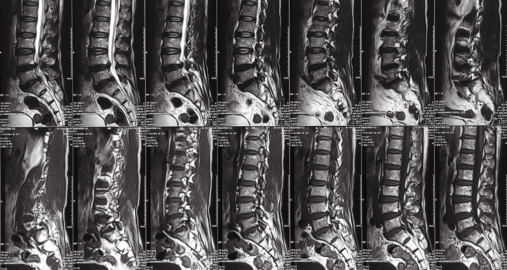
Diagnosis of diseases such as degenerative disc disease, disc herniation, assessment of spinal cord compression, nerve roots. Evaluation of anatomical abnormalities, congenital pathologies related to the lumbar spine Diagnose other problems such as spinal tumors, diagnose spinal metastases at an early stage. Diagnosis of diseases in the spinal canal such as tumors in the spinal canal, hematomas. Diagnosis of spinal diseases such as myeloma, myelitis, white matter disease

Kết quả chụp cộng hưởng từ cột sống thắt lưng
4. Notes when taking magnetic resonance imaging of the lumbar spine
MRI is a non-invasive imaging tool, so it generally does not harm the patient's body. To ensure safety and prevent complications, before conducting magnetic resonance imaging of the lumbar spine, patients should be screened so as not to violate contraindications, namely:
Patients with cardiac equipment permanently placed vessels in the body such as pacemakers, defibrillators, artificial heart valves ... or have metal fragments, metal implants in the body such as shrapnel, blood vessel clips, machines hearing aids, automatic drug pumping devices under the skin... People with psychological syndromes are afraid of closed spaces.

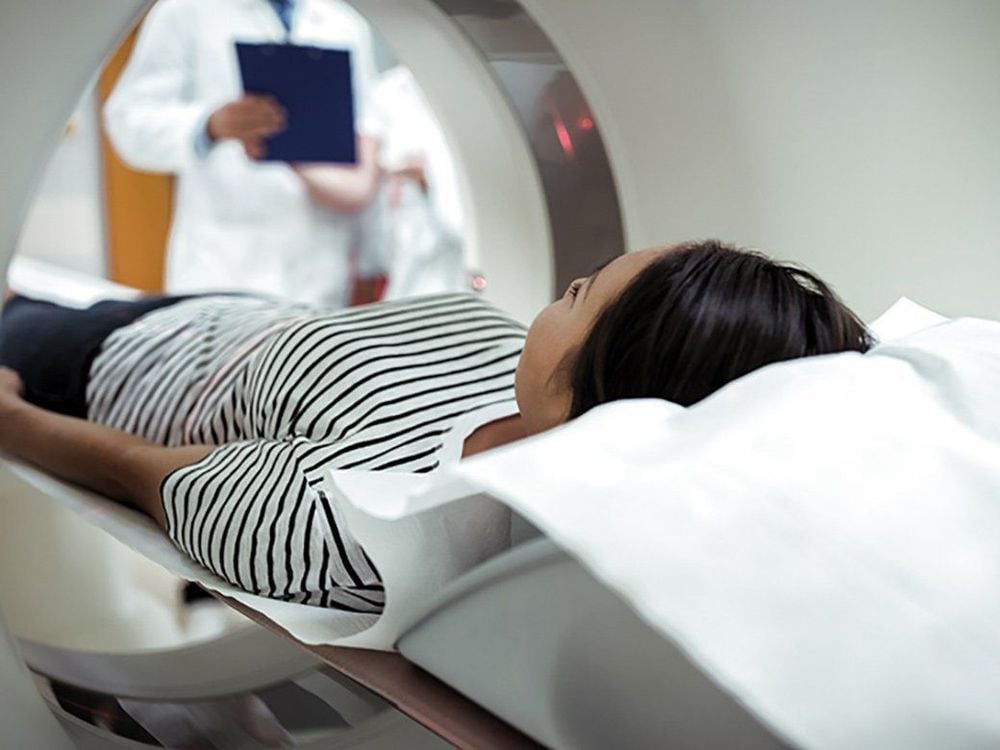
Bệnh nhân chụp cộng hưởng từ
People with severe obesity, size or body weight are too large to fit the cage. Patients do not need to fast during magnetic resonance imaging, can change into simple clothes, do not bring anything on their body and arrange to lie neatly on the plane of the machine, slowly put into the imaging cage. Meanwhile, the patient can use headphones, both to reduce noise and to be a means of communication with the outside technician; At the same time, helping the patient to relax mentally. Relax the body, relax the spirit, keep the whole body in a fixed position, the new image will appear clear, avoid stress, movement that affects the quality of image acquisition. To register for examination and treatment at Vinmec International General Hospital, you can contact Vinmec Health System nationwide, or register online HERE
SEE MORE
First time in Southeast Asia , Vinmec uses a magnetic resonance imaging machine with Silent technology Does magnetic resonance imaging (MRI) have any effect on the brain? MRI scan - the "golden" method to accurately diagnose disc herniation


