This is an automatically translated article.
The article is professionally consulted by MSc, BS. Dang Manh Cuong - Doctor of Radiology - Department of Diagnostic Imaging - Vinmec Central Park International General Hospital. The doctor has over 18 years of experience in the field of ultrasound - diagnostic imaging.Visceral CT scan for tumor hematology is a modern imaging technique that allows the detection of vascular proliferation in tumor pathologies. Tumor hematopoietic survey results are the basis for monitoring and treating tumor pathology.
1. Overview of CT scan of organs to study tumor hemodynamics
Computed tomography (CT) scan of the tumor is a technique that is integrated into the CT scan to collect cancer pathological imaging information to look for signs of angiogenesis in the tumor.Visceral CT scan to examine tumor hematology reflects vascular status in the tumor, helps in diagnosis and risk assessment, and is the basis for post-treatment follow-up of tumor pathology.
2. Indications for CT scan of organs to investigate tumor hematology
Visceral CT scan for tumor hematoma is indicated in the following cases:Thoracic tumor: mediastinal tumor, lung tumor, ... Abdominal tumor: liver tumor, biliary tract tumor, pancreatic tumor, gastroduodenal tumor colon, mesenteric tumor, spleen tumor, ... Tumor in the pelvic region: uterine tumor, ovarian tumor, bladder tumor, prostate tumor, subperitoneal cavity tumor, ...

3. Procedure for taking CT scan of organs for hemodynamic survey of tumors
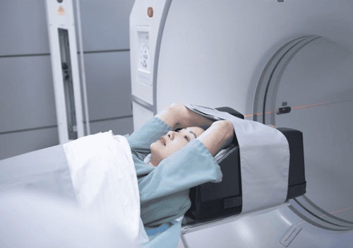
The procedure for taking a CT scan of organs for tumor hemodynamics survey includes the following steps:Step 1: Instruct the patient to lie supine on the imaging table, with both hands raised above the head to avoid image noise. Step 2: Before injecting contrast material, take a picture of the entire abdomen from the thorax down to the pelvic region, depending on the position of the tumor at the end of the exhalation. Step 3: Initial assessment of tumor characteristics in terms of density, location, size, ... At the tumor site with the largest diameter, select focal slices to continue performing CT scan later. when injected. Step 4: Inject iodinated contrast agent with dose of 40 - 70ml depending on the organ examined, the injection speed should be 6ml/s. Step 5: After contrast injection, conduct a CT scan of the viscera in the selected area. Ask the patient to hold his breath at the end of expiration to capture at a speed of 1s for 1 cut, slice thickness is 510mm. This step lasts from 25 to 30 seconds in one breath. Step 6: Transfer image data to computer, use software to measure and map perfusion, build enhancement chart of tumor lesion.

Please dial HOTLINE for more information or register for an appointment HERE. Download MyVinmec app to make appointments faster and to manage your bookings easily.


