This is an automatically translated article.
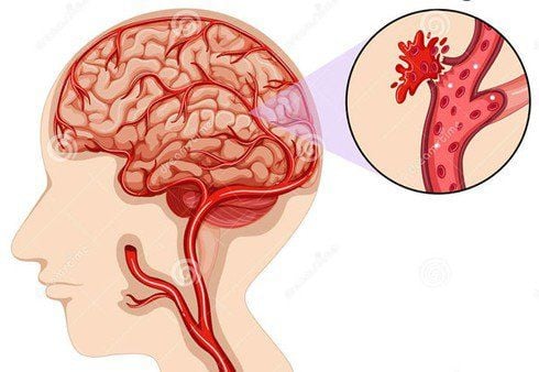
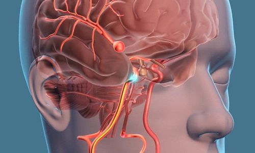
Microsurgical clamping of cerebral aneurysms is a surgery performed to treat aneurysms. When a brain aneurysm forms, it can become so thin that it is very fragile, causing bleeding to overflow into the brain parenchyma. However, although microsurgical clamping of cerebral aneurysms reduces the occurrence of this event, they still cannot completely avoid certain complications.
1. What is microsurgical clamping of cerebral aneurysms?
Clamping the brain aneurysm is a surgery that uses a clip to cut off blood flow to the brain aneurysm. This is to help prevent the aneurysm from bursting and causing a brain hemorrhage.
To perform, the patient will be sawed by the neurosurgeon to open the skull cap, guide the blood vessels to find the aneurysm. Here, highly inert metal clips such as titanium will be placed at the neck of the bag and last forever.
In case of unruptured aneurysm, proactive clamping will help prevent this risk. In contrast, in the case of a ruptured aneurysm, the patient still has an indication for clipping surgery to minimize the possibility of recurrent aneurysm rupture. However, regardless of the indications in the situation, similar to other surgical interventions, along with the benefits, microsurgical clamping of cerebral aneurysms can still cause some of the most complications. determined.

Phình động mạch não là căn bệnh được nguy hiểm cần được tiến hành phẫu thuật
2. What are the procedures to prepare before performing microsurgery to clamp the brain aneurysm?
In order to help the surgery go smoothly and minimize the possible risks, the patient should be carefully prepared for surgery with the following requirements:
General health examination Blood tests Imaging tests : ultrasound , computed tomography , magnetic resonance imaging or cerebral angiography Reporting of current medications, including prescription, over-the-counter and herbal supplements Reporting of acute illnesses recent or chronic medical conditions, previous surgery, drug allergies.. Discuss risks and benefits of treatment decisions, sign informed consent
3. How is the microsurgical clamping of cerebral aneurysms performed?
The doctor will perform craniotomy by removing a small part of the skull to gain access to the brain parenchyma. Magnetic resonance imaging right on the operating table along with microscopic observations can help doctors locate brain aneurysms.
Here, the aneurysms will be removed from the surrounding connective tissue, especially the neck area to achieve the clip here. Clamping clips are of a highly inert metal nature such as titanium to clamp the entire aneurysm, separating them from the cerebral circulation.
After checking the position of the clip is correct, the doctor will close the skull cap and the clips will stay in place permanently, preventing the possibility of rupture of the aneurysm and the occurrence of brain hemorrhage in the future .
To end the surgery, the doctor will sew up the scalp and put a bandage on the wound. This whole process takes about 3 to 5 hours. Patients need to be monitored inpatient for 4 to 6 days before being discharged from work with instructions on how to care for the incision at home and regular follow-up appointments.
4. What are the most common complications in endoscopic microsurgical clamping of cerebral aneurysms?
Immediately after the surgery ends, the patient will be removed from the breathing tube, transferred to the neuro-resuscitation department to continue monitoring vital signs until fully awake.
Next, during inpatient follow-up as well as when discharged from the hospital, the patient always needs to be monitored for the condition of the incision, the recovery of nerve function as well as for early detection of the most common complications. In endoscopic microsurgery clamping cerebral aneurysms :
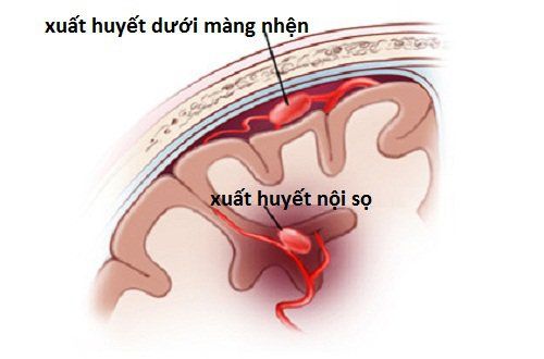
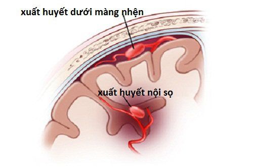
Infection: Infections of superficial wounds or deeper infections including meningitis, encephalitis or encephalitis. Hemorrhage: Bleeding can be shallow or deep as it approaches the aneurysm, causing an intracranial hematoma and stroke-like symptoms such as weakness, numbness on one side of the body, and speech disturbances. Seizures: Patients develop seizures, lose consciousness, and may require intravenous or oral medications for long-term control. Brain sequelae: Permanent nerve damage in the form of weakness, numbness, and paralysis as symptoms of a stroke. Cognitive decline: Manifestations vary, can be just subtle changes in personality, memory and thought processing... to drowsiness, lethargy, confusion and coma. Hydrocephalus or hydrocephalus: CSF circulation is disrupted, which can be temporary or permanent and requires a second surgical repair. Leakage of cerebrospinal fluid through surgery Decrease or loss of sense of smell due to leakage of CSF through the nasal sinuses Decrease or loss of vision or double vision. Acute blood loss and the need for blood transfusion during or after surgery Complications related to anesthesia, mechanical ventilation Coma and death

Một trong những tai biến nguy hiểm của nội soi vi phẫu kẹp túi phình động mạch não là hôn mê
Because surgery to clamp cerebral aneurysm is a classic intervention in the treatment of this pathology, the complications listed above occur at a very low rate. However, in cases where the patient has an indication, these risks cannot become a barrier to early intervention for the patient. However, these are also things that need to be monitored in the postoperative days, detected early and corrected in time, in order to improve the quality of surgery.
In summary, the risk of hemorrhagic stroke due to rupture of an aneurysm is improved with microsurgical clamping of the cerebral aneurysm. However, similar to other surgical interventions, this technique also contains unwanted complications. Therefore, a thorough understanding of the benefits and risks of the interventions outlined above is essential to underpin treatment decisions for this pathology.
To register for examination and treatment at Vinmec International General Hospital, you can contact Vinmec Health System nationwide, or register online HERE
Reference sources: uvahealth.com, svphm .org.au, sciencedirect.com
SEE MORE
Learn the method of plugging brain aneurysms at Vinmec Stroke: Causes, signs, and ways to avoid Brain infarction treatment methods


