This is an automatically translated article.
Article written by Th.S, BS. Ton Nu Tra My, Department of Diagnostic Imaging, Vinmec Central Park International General Hospital
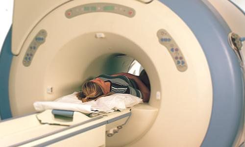
Cranial magnetic resonance imaging provides a better and more detailed assessment of brain structures than other imaging methods, and can also provide functional information (fMRI) about regions of the brain. brain activity. In particular, cranial magnetic resonance can detect cerebral infarction at an early stage, assess cerebral perfusion, thereby helping to diagnose early and intervene in time to save life in areas of brain tissue that can still be recovered.
1. What is cranial magnetic resonance?
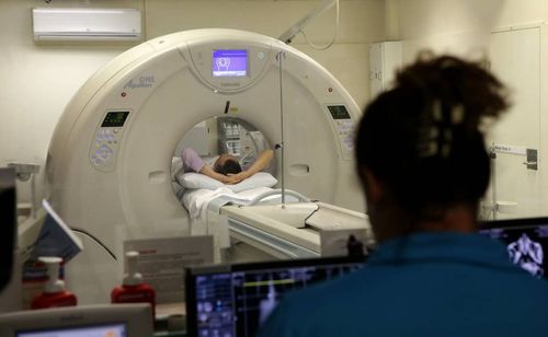
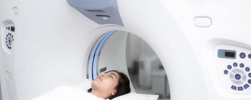
Cranial magnetic resonance (MRI) is a technique that uses strong magnetic fields, radio waves, and computers to create detailed images of the structure of the brain and skull. This method helps to detect most craniocerebral diseases such as: congenital brain defects, inflammation, stroke (cerebral infarction, cerebral hemorrhage), brain tumor, some chronic diseases such as multiple sclerosis, bleeding in patients with trauma, eye-inner ear abnormalities, pituitary abnormalities, blood vessel problems, etc. In some cases, your doctor may order an intravenous contrast agent to be injected. Better assessment of the nature of the lesions, helping to narrow the diagnosis to the exclusion of other causes based on magnetic contrast capture properties.
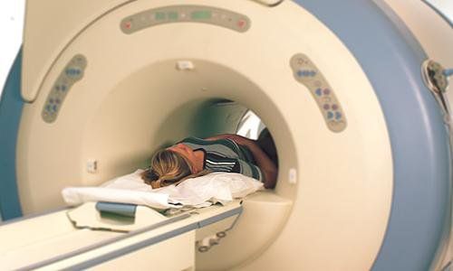
Chụp cộng hưởng từ cho phép chẩn đoán hình ảnh rõ ràng các bệnh lý liên quan đến sọ não
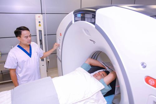
Inform your doctor about any medical problems you have, any history of surgery or allergies, and the possibility that you may be pregnant. Magnetic fields are not harmful, but they can cause some medical devices to malfunction. So you should always tell your technician or doctor if you have any equipment or metal in your body. You should leave jewelry at home. You will be asked to wear a hospital gown while performing this technique. If you suffer from agoraphobia or anxiety, you should talk to your doctor about possible sedation before the MRI.
2. The importance of cranial magnetic resonance
Cranial magnetic resonance imaging evaluates brain structures in better and more detail than other imaging methods, and can also provide functional information (fMRI) about areas of activity of the brain. In particular, cranial magnetic resonance can detect cerebral infarction at an early stage, assess cerebral perfusion, thereby helping to diagnose early and intervene in time to save life in areas of brain tissue that can still be recovered.
In addition, magnetic resonance angiography (MRA) provides detailed images of the blood vessels in the brain without the use of contrast agent (See the MRA page for more information).
Cranial magnetic resonance is commonly used in the following cases:
Birth defects of the brain; Look for causes of seizures (convulsions); Chronic headache; Suspected encephalitis, meningoencephalitis; Stroke; Brain tumors; Certain chronic diseases, such as multiple sclerosis...; Bleeding in trauma patients; Eye and inner ear disorders; Disorders of the pituitary gland; Blood vessel problems, such as an aneurysm (a balloon-like dilation of a blood vessel), an artery blockage (blockage), or venous thrombosis (a blood clot in a vein).

Bệnh nhân u não được chỉ định chụp MRI sọ não
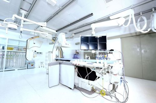
Vinmec International General Hospital put into use the magnetic resonance imaging machine 3.0 Tesla Silent technology. Magnetic resonance imaging machine 3.0 Tesla with Silent technology of GE Healthcare (USA).
Silent technology is especially beneficial for patients who are children, the elderly, weak health patients and patients undergoing surgery. Limiting noise, creating comfort and reducing stress for customers during the shooting process, helping to capture better quality images and shorten the shooting time. Magnetic resonance imaging technology is the technology applied in the most popular and safest imaging method today because of its accuracy, non-invasiveness and non-X-ray use. Customers can go directly to the system. Vinmec Health System nationwide to visit or contact the hotline HERE for support.


