This is an automatically translated article.
The article is expertly consulted by Master, Doctor Hoang Van Lan Duc - Doctor of Radiology - Department of Diagnostic Imaging and Nuclear Medicine - Vinmec Times City International General Hospital.Cerebral angiography is an advanced imaging technique widely applied in the diagnosis of cerebrovascular diseases. This is a non-invasive method of high value in disease diagnosis and early detection in acute cases, thereby providing timely treatment.
1. What is cerebral vascular computed tomography?
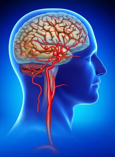
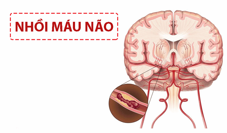
Cerebral angiography is a non-invasive imaging method that helps examine the blood vessels of the brain. By injecting a dose of contrast material into the blood, then using a scanner to capture images of blood vessels and reconstructing that image with 3-D imaging software on a computer.This is a widely used method in diagnosing cerebrovascular diseases, cerebral vascular computed tomography can diagnose diseases such as cerebral vascular malformations, cerebral aneurysms, aneurysms. Brain rupture causing cerebral hemorrhage, cavernous carotid artery bypass, or cases of cerebral infarction due to occlusion of cerebral arteries or venous sinuses,...
2. Indications and contraindications of cerebral vascular computed tomography
Indications for cerebral vascular computed tomography:Suspected cases of cerebrovascular abnormalities such as subarachnoid hemorrhage, cerebral parenchymal bleeding, intraventricular bleeding... Cerebral vascular malformations, suspected epilepsy Cerebral vascular malformation Stroke ischemic stroke. Dural arteriovenous fistula, cavernous carotid artery shunt.
In the examination area (cranium) there are many metals that interfere with the image (relative contraindication for conventional computed tomography machines). Pregnant patients (relative contraindications). Patients with contraindications to using iodinated contrast agents such as a history of allergy to contrast dyes in the past, renal failure, severe liver failure, uncontrolled hyperthyroidism, etc.
3. Cerebral vascular computed tomography scan
3.1 Preparation of Performer: A physician and radiologist is required.Means used to take computed tomography of cerebral blood vessels include:
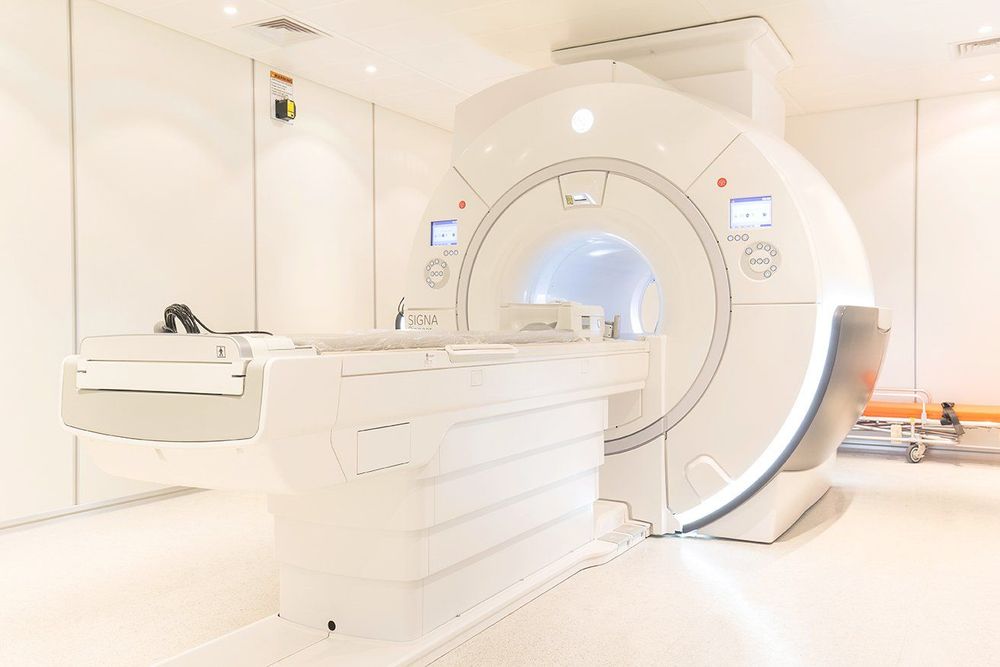
Multi-segment computed tomography machine, contrast agent pump; Film, printers and image storage systems; Water-soluble iodinated contrast agent; Supplies: 10, 20 and 50ml syringes and needles; threaded needle 18-20G; syringe for contrast injection; antiseptic solution for skin and mucous membranes; physiological saline, gloves, masks, tray sets, cotton swabs; Medicine box and necessary tools to manage complications in case of abnormalities when contrast is injected.
The patient is clearly explained about how to take the scan and the possible complications when injecting contrast material to take pictures so that he can coordinate with the radiographer; Remove items such as earrings, necklaces, hairpins... (if any); Need to fast, drink before 4 hours. Can drink water but not more than 50ml; The patient is too excited, can't lie still: Give sedation before the scan. 3.2 Procedure steps
Patient position: Lie on your back on the table, move the table into the machine with the light beam positioning for the examination area.
Shooting techniques:
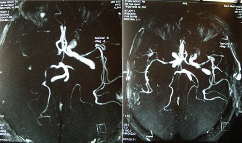
Positioning capture; Set the cranial scan field according to the schedule for the upper and lower areas of the tent; Conduct X-ray emission and image processing to evaluate brain parenchyma obtained on the workstation screen, select necessary images to reveal pathology for film printing; Place an vein with an 18G needle, connect a double-bore electric injection pump (1 barrel of medicine, 1 barrel of physiological saline). The amount of contrast medium used is usually about 1-2 ml/kg body weight; Contrast-free imaging to clear the background; Perform a bolus test of the common carotid artery at the level of the C4 cervical vertebra; Select the time to take X-rays during the injection period, set the imaging field from C4 to the top of the skull; Contrast injection and imaging (with contrast contrast with physiological saline); The acquired images will be visualized with the cerebral artery system to reveal the pathology by the MIP, VRT, and MPR programs. Evaluation of results:
Standard of photography: The image obtained should be clear, free from vibration and noise caused by movement. Displays the cerebral artery system from the base of the skull to the dome of the skull. The doctor reads the lesion, describes the lesion, and prints the results.
Some complications may occur due to contrast injection such as:
Anaphylaxis: After injection, within about 1 hour, symptoms such as itching appear. , urticaria, rash, angioedema, vomiting, bronchospasm causing difficulty breathing, stridor, laryngeal edema causing difficulty breathing, chest indrawing, hypotension, shock, the patient may lose consciousness. It is necessary to treat according to the anti-anaphylactic regimen of the Ministry of Health. Contrast-induced renal failure: A condition in which acute renal failure or increased renal failure occurs after contrast media use, in which other causes affecting renal function should be excluded. Usually occurs within about 3 days of using the contrast agent. Manifested as an elevation of serum creatinine above 25% or 44 μmol/l (0.5 mg/dl) Thyroid storm: A severe, life-threatening condition in patients with underlying hyperthyroidism. Therefore, it is very important to declare a medical history before taking a thyroid scan. Because of the risk of harm to the fetus, pregnant women, especially those in the first 3 months of pregnancy, need to consider carefully before taking the scan and often wear a lead shirt to protect the abdomen. Some people experience an allergic-like reaction, such as mild itching, but these usually go away quickly.
Patients should note that when taking a CT scan of the brain, they need to inform the doctor about their illness, pregnancy, etc. To avoid the risk of complications due to the use of X-rays for imaging and the use of contrast agents. .
Currently, Vinmec International General Hospital is equipped with 128 and 256-layer CT scanners capable of supporting the best diagnosis with fast speed allowing one rotation to capture the entire heart, helping to make optimal diagnosis. coronary, vascular and systemic. In particular, this machine can reduce up to 90% of radiation dose, so it can be taken for pregnant women when indicated.
Master. Doctor Hoang Van Lan Duc was formally trained and graduated with a Master's degree from Hanoi Medical University. The doctor has more than 10 years of experience in the field of imaging, especially in diagnosing with emergency internal, surgical, abdominal, thoracic, musculoskeletal, neurological and thyroid, breast, ...
Please dial HOTLINE for more information or register for an appointment HERE. Download MyVinmec app to make appointments faster and to manage your bookings easily.


