This is an automatically translated article.
The article is professionally consulted by Master, Doctor Tong Van Hoan - Emergency Medicine Doctor - Emergency Department - Vinmec Danang International Hospital.The ventilator helps the patient to breathe in case the patient cannot breathe on his own for any reason (respiratory failure due to pneumonia, myasthenia gravis, effusion, hemothorax ..). Although mechanical ventilation has many benefits such as providing oxygen to the patient, the patient has to face a number of complications such as prolonged respiratory infection, malnutrition, electrolyte disturbances, pressure ulcers caused by prolonged lying down. ... Therefore, taking care of ventilator patients plays an important role in preventing complications later on.
1. What is mechanical ventilation?
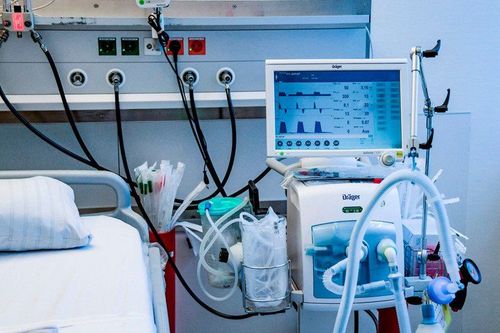
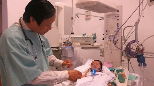
What is a ventilator? Mechanical ventilation, also known as mechanical ventilation (English name is mechanical ventilato) is used for treatment and life support. Ventilators are used when the patient is unable to breathe on their own. Most people need support from a ventilator because of a serious illness that requires care in a hospital or intensive care unit (ICU).Patients with ventilators will need to be treated and cared for for a longer period of time in Health facilities such as hospitals, rehabilitation facilities or home care.
Ventilators are used to:
Bring oxygen to the lungs and body; Helps the body remove CO2 from the lungs; Reducing effort for breathing in patients who have difficulty breathing on their own or have medical problems that prevent the patient from breathing on their own, then the patient will have difficulty breathing and discomfort; Helping someone who can't breathe because of an injury to the nervous system, such as the brain or spinal cord, or who has paralysis of the respiratory muscles
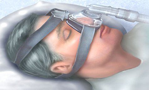
Máy thở có tác dụng đưa oxy vào phổi và loại bỏ CO2
2. How many types of ventilators are there?
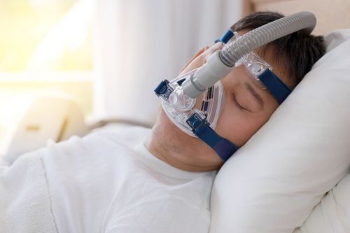
How many types of ventilators are there? There are two types of mechanical ventilation:Invasive mechanical ventilation: mechanical ventilation through the endotracheal tube or a tracheostomy. Non-invasive ventilation: mechanical ventilation through a nasal mask or a nose and mouth mask.
3. Benefits of mechanical ventilation
The main benefits of mechanical ventilation are as follows:The patient does not have to spend energy to breathe, so the patient's respiratory muscles have time to rest; The patient's body has time to recover; Help the patient get enough oxygen and get rid of carbon dioxide; Protects the respiratory tract and prevents damage from aspiration of gastric juices. However, it should be noted that mechanical ventilation does not heal the patient's illness. Instead, mechanical ventilation allows the patient to stabilize while the doctor uses drugs and other treatments to treat the illness or injury.
Usually, as soon as the patient can breathe effectively on their own, they are removed from the ventilator. Caregivers will perform a series of tests to check the patient's ability to breathe on their own. When the cause of the breathing problem improves and it is felt that the patient can breathe effectively on their own, they are removed from the ventilator.
4. Risks of mechanical ventilation
The main risk of mechanical ventilation is infection, because invasive ventilation uses a breathing tube inserted into the patient's lungs, allowing bacteria to enter the lungs. This risk of infection increases with the length of time the patient is on the ventilator and peaks at about two weeks.Another risk is lung damage from overuse or from mechanical ventilation that ruptures the alveoli of the lungs. Sometimes, the patient cannot be weaned off the ventilator and needs more prolonged breathing support.
In some cases, the patient will be removed from the mouth and have to open the trachea to support breathing from the trachea. Prolonged use of a ventilator increases the risk of death if the patient is not weaned off the ventilator early.
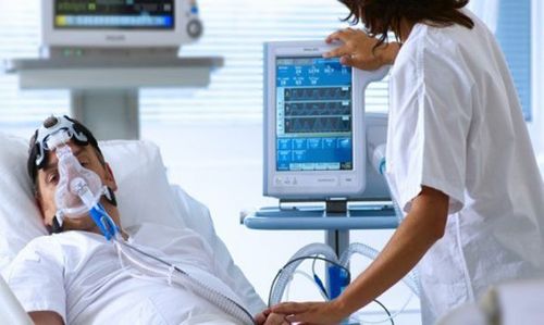
Thở máy sẽ có nguy cơ bị nhiễm trùng phổi
5. Caring for ventilator patients
5.1. Caring for an endotracheal tube or tracheostomy5.1.1. Objective
Intubation or tracheostomy must be ventilated. Ensure that the endotracheal or tracheostomy position is in the correct position. Avoid infection 5.1.2. Perform techniques
Clearing the airways by flapping technique, sputum suction technique (see technical procedure of vibrating for respiratory care). Perform the technique of changing the dressing of the tracheostomy tube, opening the trachea according to the correct procedure to ensure the correct position to avoid infection. Check endotracheal cuff pressure, tracheostomy (see endotracheal care, tracheostomy). 5.2. Caring for the Patient Non-invasive breathing through a nose-mouth mask
The size of the mask must fit the patient's face. When fixing the mask, it should not be too tight, which can easily cause pressure ulcers (nose bridge) or too loose, causing air to leak out, reducing airway pressure. Fix the mask: the top loops over the head above the ears, the bottom loops around the back of the neck. The device can be turned off when the patient coughs up phlegm. Remove the non-invasive ventilator when the patient eats, drinks (otherwise it will cause aspiration of food and water into the lungs), or eats and drinks through a nasogastric tube. Must explain to the patient to cooperate, and unwanted effects (stomach bloating, feeling of suffocation...). 5.3. Ventilator Activity Monitoring Care
5.3.1. Ventilator supplies
Power: always plugged into the mains. When power is on, the AC indicator will light up. It has the effect of both running the ventilator and charging the battery of the machine so that in the event of a power failure, the machine will automatically switch to battery power (the battery life lasts depending on the type of ventilator). Oxygen source: connected to the oxygen supply system, when the machine is turned on, there will be no oxygen pressure alarm (O2 Pressure) Compressed air source: connected to the compressed air supply system, when the machine is turned on, there will be no pressure alarm compressed air force (compressor). 5.3.2. Air duct system
The tubes that carry air in and out of the patient must always be lower than the endotracheal tube (tracheostomy) to avoid standing water on the wall of the tube entering the endotracheal tube (tracheostomy) causing pulmonary aspiration. . Replace the airway (ventilator wire, T-wire) when there is a lot of sputum or the patient's blood in the air duct. There must always be a water trap on the inlet and outlet pipes (water standing on the wall of the pipe will flow into this water trap, so the water trap is set to the lowest position). Pay attention to pour standing water in the water trap cup, if it is full, it will obstruct the airway and there is a risk of water flowing into the lungs. 3. Airway Humidification System
This system is located in the inlet airway, before air is introduced into the patient. The humidifier uses distilled water, must ensure that the water level in the tank is always within the allowable limit. Burner of the humidification system: 30 - 370C. Has the effect of increasing the humidity of the inhaled air, thus avoiding the phenomenon of dry sputum causing obstruction. The higher the combustion temperature, the faster the evaporation rate of the water in the humidifier, so it is necessary to regularly add water to the humidifier. With a temperature of 350°C, 2000ml/day is used up. Some ventilators have additional heating coils located in the inlet and combustion chambers of the humidification system. Therefore, the wire used for this type of ventilator must also have a heat-resistant effect. 5.3.4. Monitor parameters on ventilator, ventilator alarm system.
6. Monitor ventilator patients
Heart rate Blood pressure SpO2 Temperature Arterial blood gases Sputum properties: abundant, cloudy (with respiratory infection) Stomach fluid. Urine (color, quantity). Other drainages: pleural, pericardial, ventricular drainage..7. Complications – treatment
7.1. To avoid reflux of gastric juice, oropharyngeal fluid into the lungsCheck the pressure of the balloon daily. Have the patient lie down with the head elevated 300 (if there are no contraindications) Give the patient a gastric drip, not more than 300 ml /meal. (according to the procedure for feeding through a nasogastric tube) When there is reflux of fluid into the lungs: postural drainage or bronchoscopy with a flexible bronchoscope. 7.2. Pneumothorax
Manifestations: Patient is cyanotic, SpO2 decreases rapidly, pulse is slow, thoracic side is full of pneumothorax, percussion echoes, subcutaneous pneumothorax... Must conduct air drainage immediately, if not open airway in time Time will make the pressure in the chest increase very quickly leading to respiratory failure and acute cardiac tamponade, the patient quickly leads to death. Perform minimal emergency pleural effusion with a large enough drainage tube Connect to a continuous suction machine with a pressure of 15-20cm H2O. The drain should be checked daily for kinks or blockages. The suction system must be tight enough, in good working order, the water in the drainage tank from the patient must be closely monitored and poured daily. The water in the bottle to detect air out must always be clean. Leave the tube to drain until the air is empty, and after 24 hours, clamp it and then take a chest X-ray to check, if satisfactory, the lungs are fully expanded -> pull out the drain tube. . 7.3. Ventilator-related pneumonia
Manifestations: cloudy sputum, appearing in many places; fast heart beat; fever or hypothermia; increased white blood cell count; Chest X-ray showed new lesions. Test bronchial fluid (fresh endoscopy, culture): to identify pathogenic bacteria. Blood culture when sepsis is suspected. Re-evaluate the processes of suctioning sputum, cleaning the wiring and ventilators to see if they are sterile. Use strong broad-spectrum antibiotics, combine antibiotics according to the protocol. 7.4. Prophylaxis of peptic ulcer: use drugs to reduce gastric secretions: proton pump inhibitors, gastric coatings.
7.5. Prevention and care of pressure sores
Change position every 3 hours: straight, right side, left side (if there are no contraindications) to avoid long-term pressure on one place. In addition to the anti-ulcer effect, it also has the effect of preventing atelectasis. If the patient is expected to lie down for a long time: put the patient on a water mattress, the air mattress will change the position of the inflatable automatically. When there is redness of the pressure area: use synaren to rub on the pressure area. When there is an ulcer: clean, cut and change the dressing of the ulcer daily. 7.6. Prophylaxis of deep vein thrombosis due to prolonged lying
Change posture, passive exercise for patients: avoid circulatory stagnation. Systematic pulse check: detect occlusion, venous or arterial occlusion Anticoagulation: Low molecular weight heparin: Lovenox, Fraxiparin. Taking care of ventilated patients is very important and directly affects the patient's health and recovery. Therefore, patients need to be cared for by a team of qualified, professional and qualified doctors, nurses, and nurses.
Vinmec International General Hospital is a medical facility with highly qualified and professional human resources from famous domestic and international medical universities. The most modern and advanced equipment system in Vietnam. Here, patients are cared for wholeheartedly, thoughtfully, with quick recovery time.
To register for examination and treatment at Vinmec International General Hospital, you can contact the nationwide Vinmec Health System hotline, or register online HERE.
Reference sources: thoracic.org, my.clevelandclinic.or
MORE:
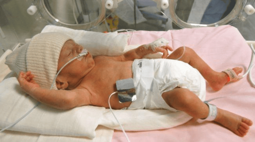
Techniques of continuous positive pressure breathing through the nose at Vinmec Phu Quoc Notes when caring for patients with ventilator-associated pneumonia Caring for mechanically-ventilated newborns


