This is an automatically translated article.
With the advancement of medicine along with the support of modern equipment such as nerve locator, the surgical implementation of cranial nerve tumors under the skull base has achieved remarkable achievements. The surgeries help patients recover faster and reduce the length of stay in the hospital.
1. What is a cranial nerve tumor at the base of the skull?
Some brain tumors at the base of the skull that patients may encounter such as:
Tumors often develop in the base of the skull anteriorly such as: ethmoid sinus tumor, frontal sinus tumor, nasolabial orbital tumor, meningioma of the olfactory groove , meningiomas on the pituitary gland, in the pituitary gland, and in the skull base frontal brain tumor. Tumor appearing at the base of the middle skull: Basal temporal meningioma, sphenoid sphenoid tumor, lateral wall tumor of the cavernous sinus, apex of the scapula, cord tumor V, tumor anterior of the scapula, tumor in the basal temporal region, tumor in the gyrus region code - inferior surface of the temporal lobe. Developments in the posterior skull base: Sliding sulcus, posterior squamous cell tumors Brain tumors below the skull base, if not treated early, will cause dangerous complications and slow recovery. Therefore, surgery for cranial nerve tumors under the skull base is an effective and successful treatment method after surgery.
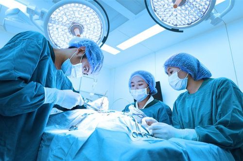
Phẫu thuật u thần kinh sọ não dưới nền sọ là phương pháp hiệu quả được áp dụng nhiều
2. Surgery for cranial nerve tumors under the skull base
Step 1: Preparation
On the side of the hospital: To perform cranial nerve surgery, the hospital needs to prepare a surgical team including: 3 doctors (1 main surgeon and 2 supporting doctors) ; 2 nurses; 1 anesthesiologist and 1 anesthesiologist. Full range of surgical instruments, including: endotracheal anesthesia equipment, conventional craniotomy kits, microsurgery glasses, nerve positioning system, consumables, closed lower drainage set Skin.... On the patient's side, the hair in the hairline area is shaved, cleaned and disinfected. In addition, the patient needs to be clinically examined such as: Magnetic resonance imaging, tests for surgery, indications for surgery and most importantly, a written commitment to surgery before surgery. Step 2: Procedure
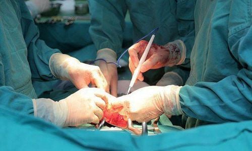
The patient is placed in a lying position depending on the location of the tumor and the way to get the tumor, can lie on his back or on his side. Conducting endotracheal anesthesia and placing a neuropositioning system The doctor made a skin incision to dissect the fascia, expose the skull, drill the skull bone, open the skull cap close to the base of the skull and open the dura, aspirate cerebrospinal fluid to flatten the brain If the patient has surgery for the tumor at the base of the skull first, it is necessary to remove the tumor from the vascular structure, cut the tumor stalk, and remove the tumor with ultrasound If the patient has surgery for the tumor at the base of the middle skull: Usually the wing needs to be ground. to reduce the sphenoid bone, in some cases, it is necessary to grind the anterior saddle to control bleeding, cut the tumor attachment point, dissect and aspirate the tumor with ultrasound If the patient has surgery for the tumor at the top of the stony bone, 2/3 above the slide : Grinding the top of the stony bone through the Kawase triangle, dissecting the peduncle, and sucking the tumor. Stop bleeding and close the dura. Step 3: End of surgery
Perform fixation of skull, balance, muscle, subcutaneous, skin. The surgery is over.

Sọ được mở một phần để thuận tiện cho quá trình phẫu thuật
3. Post-surgery
The operation is determined to be successful only if there are no postoperative complications.
Therefore, the process of monitoring and dealing with accidents should be done closely. Patients need to be treated and monitored in the recovery room after surgery to wean off the machine and remove the endotracheal tube. Especially monitor incision bleeding, mental status, convulsions
Some complications the patient may encounter such as:
Bleeding. If the bleeding is small, the patient can be treated with internal medicine. If there is a lot of bleeding, you will need to have surgery again. Ventricular dilation: Drain the ventricles out. CSF fistula through incision: lumbar puncture to drain CSF to relieve pressure and medication or surgery to close the dural opening. Progressive cerebral edema: should be treated according to the cause. The patient was prescribed tomography to find the cause. Any questions that need to be answered by a specialist doctor as well as customers wishing to be examined and treated at Vinmec International General Hospital, please book an appointment on the website to be served.
Please dial HOTLINE for more information or register for an appointment HERE. Download MyVinmec app to make appointments faster and to manage your bookings easily.