This is an automatically translated article.
Cerebellar tumors are tumors arising from the cerebellum region and the fourth ventricle in the posterior fossa region. The disease will seriously affect the health as well as the spirit of the patient. Open the skull cap surgery for cerebellar tumors is the treatment of choice with high efficiency.
1. What is cerebellar tumor?
The pupal lobe, cerebellar hemisphere, and fourth ventricle are the sites where the tumors arise. Cerebellar tumors include:medulloblastoma, astrocytoma, ependymoma, and some other less common tumors. Children are the most susceptible to cerebellar tumors. The cause of brain tumors in children is thought to be cerebellum development is not clear; Some cases are hereditary.
Cerebellar tumors cause mild to severe symptoms depending on the condition and location of the tumor. Mild headache (this is the cause of increased cranial pressure), vomiting, papilledema, back pain... In severe cases, the head is large, the fontanel is wide, and the skull joints are dilated. This symptom occurs mostly in children. In addition, the patient will also experience symptoms such as: Language disorder, lower limb movement, limb tremor, ...
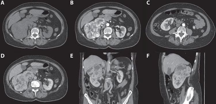
To diagnose the disease, doctors often appoint diagnostic imaging such as: Magnetic resonance imaging, taking layers,... Diagnostic imaging will help the doctor probe as well as determine the location, condition, and size of the tumor.
In the treatment of cerebellar tumors, opening the skull cap is a highly effective treatment method.
Before the surgery, the patient is explained in detail about the before and after surgery, as well as a written commitment to the surgery from the patient's family.

Chụp cắt lớp giúp chẩn đoán u tiểu não
2. Open the skull cap for cerebellar tumor surgery
Step 1: Preparation
To perform craniotomy, the hospital needs a highly skilled and experienced surgical team including: 1 main surgeon + 2 auxiliary surgeons; 2 nurses; 1 anesthesiologist and 1 anesthesiologist.
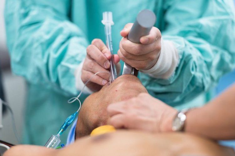
Full surgical instruments including: Instruments and endotracheal anesthetics; Conventional craniotomy kits; Microsurgical glasses; Consumables and Sealed Drains placed under the skin.
Patient is shaved clean, cleaned and disinfected the surgical area.
Step 2: Perform surgery
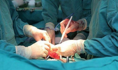
Put the patient in a prone position, good crouching, endotracheal anesthesia Install a nerve positioning system (if necessary, especially in cases where the tumor is similar to white matter or tumor below the parenchyma of the cerebral cortex). Make an incision in the midline below the occipital line, or the line below the occipital line. The fascia was then dissected, exposing the skull. Next, the skull will be drilled, opened the lower occipital skull, under the transverse sinus and aspirated cerebrospinal fluid to deflate the brain. In case the patient has a tumor in the cerebellar hemisphere, the doctor will use a suction machine or an ultrasound knife to open the cerebellar cortex and dissect the tumor. If the patient has a tumor in the lobule, use an ultrasound knife to dissect around the tumor to control the source of bleeding. The last step is to stop bleeding and close the dura, fix the skull. Step 3: Finish surgery

Bệnh nhân cần gây mê nội khí quản trước khi phẫu thuật
3. Post-surgery
After surgery, the patient will be transferred to the recovery room for monitoring and extubation.
Like other surgeries, opening the skull cap for cerebellar tumor surgery as well as encountering some complications such as: Bleeding, dilated ventricles, progressive cerebral edema,... Therefore, patients need be monitored, detected and dealt with quickly.
Any questions that need to be answered by a specialist doctor as well as if you have a need for examination and treatment at Vinmec International General Hospital, please book an appointment on the website to be served.
Please dial HOTLINE for more information or register for an appointment HERE. Download MyVinmec app to make appointments faster and to manage your bookings easily.


