This is an automatically translated article.
The article was written by Doctor Hoang Thi Thu Huong - Ultrasound Doctor, Obstetrics Department - Vinmec Times City International General Hospital.
The placenta develops from the trophoblast layer of the embryoblast around day 6 after fertilization. The placenta is first noticed on ultrasound as an area of thick echogenicity around 9-10 weeks gestation. The placental end of pregnancy has a diameter of 20cm and a weight of 400-600 g. The thickness of the placenta corresponds to the number of weeks of gestation in mm.
1. Diagnosis of placenta previa
The placenta connects to the baby through the umbilical cord. In most pregnancies, the placenta attaches to the bottom or side of the uterus.
Placenta previa is the placenta partially or completely attached to the lower uterine segment. It is one of the most common causes of bleeding in the second half of pregnancy. In the United States, the frequency of placenta previa in term pregnancies is 4.8/1000 births.
The first symptom of placenta previa may be bleeding, so early diagnosis and proactive cesarean delivery should be planned if placenta previa persists into the third trimester of pregnancy.
2. Risk factors of placenta previa

Trước đó đã từng phá thai có nguy cơ bị rau tiền đạo
History of previous cesarean section. Abortion, previous uterine surgery. The mother smokes, is older, gives birth many times, uses stimulants. Multiple pregnancy.
3. Diagnosis and classification of placenta previa
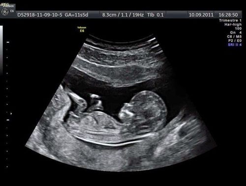
Only detected by ultrasound to locate the placenta
Over 16 weeks + The edge of the placenta is > 2cm from the inner hole: normal
+ The edge of the placenta is less than 2cm from the inner hole of the cervix, do not cover the inner hole of the cervix. uterine: low placenta, transvaginal ultrasound follow-up at 32 weeks is recommended.
32 weeks + Placental margin < 2cm, covering the hole in the cervix: placenta previa, ultrasound at 36 weeks with a transvaginal probe.
+ If there is bleeding, appoint an earlier follow-up ultrasound.
+ Color Doppler ultrasound with transvaginal transducer at 32 weeks is recommended to rule out vagal previa, assess placental perfusion, and assess risk of placenta accreta.
4. Manage striker vegetables

Rau tiền đạo
With the diagnosis of placenta previa, the patient is scheduled to deliver by elective caesarean section at 36-37 weeks. However, some patients have complications and require emergency cesarean section at early gestational age. more if there is bleeding.
Vinmec International General Hospital offers a Package Maternity Care Program for pregnant women right from the first months of pregnancy with a full range of antenatal care visits, periodical 3D and 4D ultrasounds and routine tests to ensure that the mother is healthy and the fetus is developing comprehensively.
Pregnant women will be consulted and checked for health under the close supervision of experienced and specialized Obstetricians, helping mothers have more knowledge to protect their health during pregnancy as well as reduce reduce complications for mother and child.
References:
Ultrasound in Obstetrics and Gynecology, a Practical Approach ncbi.nlm.nih.gov
Please dial HOTLINE for more information or register for an appointment HERE. Download MyVinmec app to make appointments faster and to manage your bookings easily.