This is an automatically translated article.
The article was professionally consulted with the Radiologist, Department of Diagnostic Imaging - Vinmec Central Park International General Hospital.Echocardiography is an advanced medical technique that helps to detect heart-related diseases, from which an effective treatment plan can be developed.
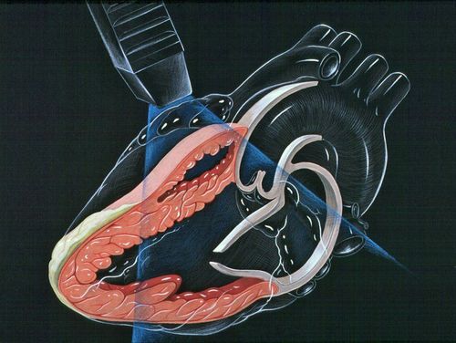
1.What is an echocardiogram? An echocardiogram is a technique that uses ultrasound waves to create images of the heart. This is a non-invasive technique that does not use radiation and usually causes no side effects. Through echocardiography, the doctor can survey and evaluate details such as:
Size and wall thickness of the heart chambers. Heart valve activity. The direction of blood flow through the heart. Areas of heart muscle tissue that are damaged or weakened. Pericardial effusion. In addition, echocardiography can also be used as a tool for routine heart health check-ups or reevaluations with patients following a heart attack or stroke.
Besides, echocardiography also helps doctors evaluate:
Assess the pumping action of the heart. Evaluation of electrical current changes in the heart (ECG). Diagnose heart disease such as cardiomyopathy, valvular regurgitation, blockage or dilated heart chambers. Locate the thrombus or tumor. Assess intracardiac pressure to diagnose pulmonary hypertension. Identification of congenital heart abnormalities in infants and young children. Monitor your heart's response to various heart treatments such as heart failure medications, artificial valves, and pacemakers. 2. Commonly used methods in echocardiography There are many methods of echocardiography available today, but below are the most common and effective methods:
Transthoracic echocardiography Transcardicardial echocardiography The chest wall is the most common echocardiographic technique. The ultrasound probe is placed on the outside of the chest near the heart. Applying gel on the chest helps the sound waves to travel better.
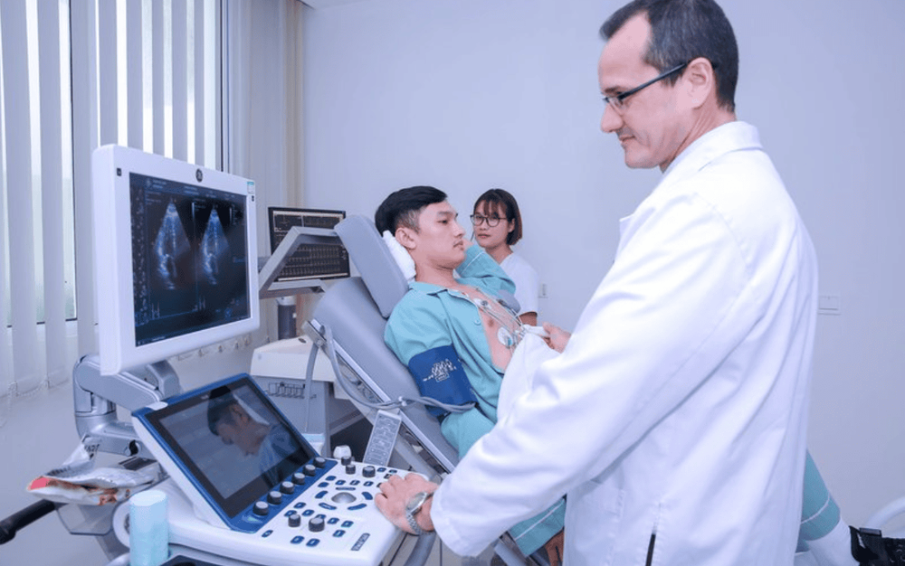
Hình ảnh siêu âm tim qua thành ngực tại Vinmec
Transesophageal echocardiography Transesophageal echocardiography uses a thinner probe attached to the end of the endoscope. This tube will be inserted into the esophagus to help the doctor better see the details of the heart from behind.
This echocardiogram will provide a more detailed picture of the heart than a traditional echocardiogram because it provides more detailed and clear images.
Doppler ultrasound Doppler ultrasound is used to check blood flow, measure pulmonary artery pressure. Your doctor may use color doppler ultrasound to map the direction and speed of blood flows in the heart.
Normally blood flowing towards the transducer appears red, bleeding out is blue.
Results of Doppler ultrasound can check, detect some heart diseases, blood vessels and evaluate cardiac output.
Three-dimensional echocardiography Three-dimensional (3D) echocardiography is the method that produces the most detailed images of the heart. This method helps doctors have a holistic view of diseases:
Evaluate heart valve function in people with heart failure Diagnose heart problems in babies and children Examine structures before heart valve replacement surgery or surgical intervention Assess the function of the heart in 3D. Reconstruction of complex structural images of the heart Stress echocardiography This method of stress testing involves exercise (usually walking or jogging on a treadmill). During the test, your doctor will monitor your heart rate, blood pressure, and electrical impulses. Doctors usually perform this ultrasound before and after exercise. This method contributes to the diagnosis of:
Ischemic heart disease Coronary heart disease Heart failure Problems affecting the heart valves Fetal echocardiogram Doctors can use fetal ultrasound to see the functioning of the heart. fetal heart. This technique usually takes place around 18-22 weeks of pregnancy.
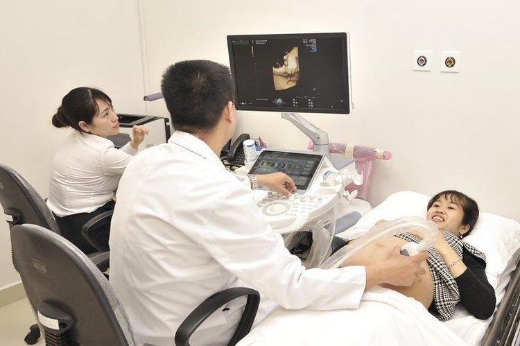
Siêu âm tim thai giúp bác sĩ sớm phát hiện bất thường tại vùng tim của thai nhi
3. When to do an echocardiogram? When you have signs of heart abnormalities, such as shortness of breath or chest pain, an echocardiogram is essential. At that time, doctors will ask you to have an echocardiogram in the following cases:
Finding abnormalities from other tests or while the doctor listens to the heart through a stethoscope. Detects cardiac signs such as heart palpitations and fluttering in the heart. Detect heart defects such as: congenital heart disease,... while working,... Suspect the patient has pulmonary hypertension. Detecting the patient has a relative with previous heart disease. 4. Benefits from echocardiography As mentioned, performing ultrasound will help doctors accurately and objectively diagnose the patient's medical condition. When using an ultrasound machine, doctors can perform an echocardiogram to look at a patient's heart. The ultrasound waves from the ultrasound machine allow doctors to see how the patient's heart is beating and pumping blood.
Through these images, the doctor can find many abnormalities in the heart muscle and heart valves.
As a widely used medical method, echocardiography helps to check for abnormalities of the heart including:
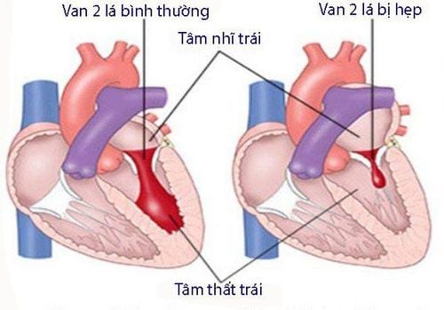
Shape, size, activity, contraction of the heart. The size and pumping process of the valve and the distance from the heart wall. The pumping power of the heart and the volume it pumps blood to other organs of the body. This pumping power is calculated per minute and averaged. Check the heartbeat. Detect abnormalities or damage to the heart such as heart valve regurgitation, wide or narrow heart valves, abnormal blood clots in the heart, problems with the outer lining of the heart. Detect abnormal tumors of unknown cause, causing compression and pain in the chest.
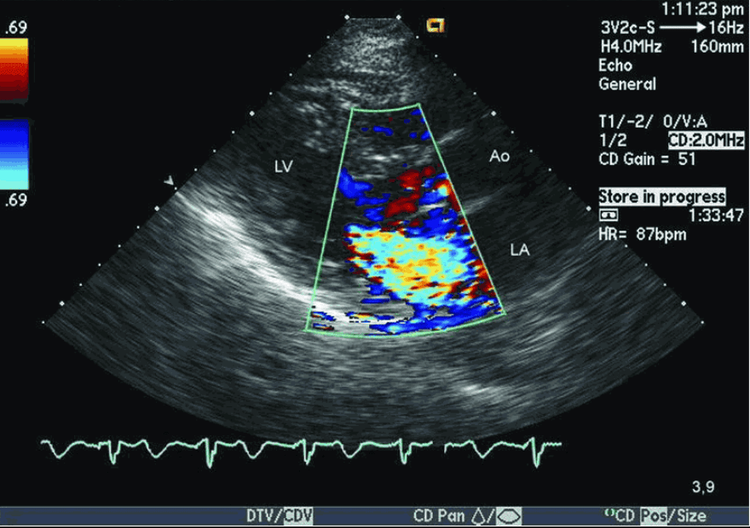
Hình ảnh sieu âm tim phát hiện bệnh lý hở van 2 lá
Echocardiography is an effective solution to help doctors diagnose as well as easily detect heart-related diseases such as heart valve regurgitation, heart failure, myocardial infarction, myocarditis,... This helps patients have time to prepare psychologically and quickly receive timely treatment to help ensure quality of health before worsening conditions occur.
5. Steps to perform an echocardiogram Before an echocardiogram Before an echocardiogram, you can eat, drink, and take medications as usual if you only had a regular echocardiogram. In cases where you have difficulty swallowing, you should inform your doctor as this can cause difficulties during a transesophageal echocardiogram.
In addition, during a transesophageal echocardiogram or an exercise echocardiogram, your doctor may ask you not to eat anything for a few hours for the most accurate echocardiogram.
During the transesophageal echocardiogram, the doctor may use sedation, so before going to the ultrasound, you should accompany your loved one to move safely.
During the ultrasound, the doctor will ask you to lie on the bed and pull the shirt up from the waist so that the doctor uses the patches that are electrodes into the body and will monitor the electrocardiogram running on the live projection screen.
The doctor will also use a gel to apply to the area to be ultrasound to increase the transmission of ultrasound waves. At this point, the transducer will be moved back and forth across your chest and record an echocardiogram of your heart. Your doctor will take pictures of the most important parts of your echocardiogram if any heart disease or restricted blood flow to the heart muscles is found.
If you have a transesophageal echocardiogram, you will be anesthetized with an inhaler or given a sedative to make the ultrasound easier.
After the echocardiogram After your echocardiogram is completed, if there are no problems, you can receive the results and go home and can go about your daily activities.
If the case is detected, the doctor will assign you to stay for treatment or prescribe medicine for you depending on your medical condition.
Echocardiography is a superior method in medicine, helping patients to detect disease early and treat it effectively. Therefore, patients need to pay attention to perform echocardiography regularly. The echocardiography process is also not complicated, so patients can rest assured to go for regular check-ups.

Định kỳ kiểm tra sức khỏe tim mạch giúp người bệnh được điều trị kịp thời
Vinmec International General Hospital is one of the hospitals that not only ensures professional quality with a team of leading medical professionals, modern equipment and technology, but also stands out for its examination and consultation services. comprehensive and professional medical consultation and treatment; civilized, polite, safe and sterile medical examination and treatment space. Customers when choosing to perform tests here can be completely assured of the accuracy of test results.
Please dial HOTLINE for more information or register for an appointment HERE. Download MyVinmec app to make appointments faster and to manage your bookings easily.


