This is an automatically translated article.
The article was professionally consulted by Specialist Doctor I Tran Cong Trinh - Radiologist - Radiology Department - Vinmec Central Park International General Hospital. The doctor has many years of experience in the field of diagnostic imaging.Hepatobiliary ultrasound is a modern imaging technique that makes an important contribution to the diagnosis and treatment of liver and biliary diseases. What are the standard slices used in the visualization of the hepatobiliary system?
1. What is hepatobiliary ultrasound?
Hepatobiliary sonography is a method that uses high-frequency sound waves to shape and reconstruct images and structures of the hepatobiliary system inside the body such as lobes, lobes, subsegments and related structures such as the veins and veins. inferior aorta, aorta, hepatic vein, portal vein. This method is applied in most medical facilities as an effective subclinical test for the diagnosis, monitoring and treatment of hepatobiliary diseases.Liver ultrasound will usually be indicated when:
Patient has the following symptoms: yellowing of the skin and eyes (hyperbilirubinemia) Abdominal distention: due to free fluid in the abdomen On the skin appear spider webs as thin as blood vessels small Urine turns dark due to the amount of bilirubin in the liver excreted along with urine Biliary ultrasound will be indicated when:
The patient's skin turns yellow for more than 2 weeks, especially for young children who have gone through the golden period physiological skin but still have signs of severe jaundice. For a long time, despite eating and living in moderation, the color of stools is proportional to the severity of biliary tract disease. Dark urine Children malnutrition, growth retardation, high degree of anemia are also some abnormal signs of bile.

2. Basic slices in hepatobiliary ultrasound
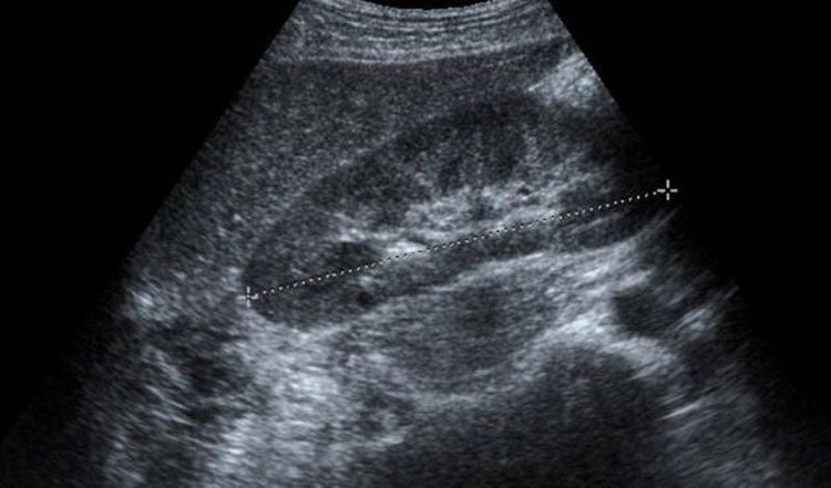
Hepatobiliary ultrasound consists of 6 basic slices, but in reality these are only the first steps in ultrasound. Depending on the patient's condition and disease condition, the technician can change the ultrasound image to be as clear as possible. The hepatobiliary ultrasound slices include:Longitudinal slices of the liver: transducer transversely transverses from front to back through the abdominal aorta to help visualize the left lobe of the liver, the 2-3 lobes, the round ligament, caudal lobe, left hepatic vein, lower segment IV, square lobe, gallbladder, portal vein, vena cava, middle hepatic vein, right hepatic vein and Morison space Left hepatic transverse section: transducer translation from from top to bottom along the axis of the left portal vein branch and cut back from below the right costal margin through the right portal vein branch to help visualize: left hepatic lobe, heart, left hepatic vein, caudal lobe, round ligament, Lower segment III and biliary tract Transverse section of the mid-liver: transducer translates from top to bottom (or vice versa) through the right liver, gallbladder and right kidney, helping to visualize the middle and anterior segments of the liver, the starting part portal vein, caudal lobe, left branch of portal vein, right and left branch of portal vein, round ligament, cystic ligament and inferior border of liver, kidney, gallbladder Right hepatic transverse section: transducer transducer From top to bottom, it helps to visualize the portal vein, lower lobe of the liver and kidney. Cross section of the gallbladder: helps to visualize the right branch of the portal vein, the venous ligament, the neck of the gallbladder, the junction of the neck and the body of the gallbladder, Gallbladder body and gallbladder fundus Longitudinal slice of gallbladder: helps to visualize vena cava, duodenum, portal vein branch, right portal vein branch, duodenum, gallbladder body, right kidney.

3. Some notes when ultrasound hepatobiliary
Before the hepatobiliary ultrasound, the patient needs to fast for 6-8 hours because after eating, it can cause the gallbladder to shrink, which interferes with the examination, leading to the omission of lesions. However, in emergency cases, the patient does not necessarily have to fast before that, but it is still possible to conduct ultrasound immediately in combination with clinical examination.For young children, because their bodies are weaker than adults, they cannot fast for 8 hours like adults. At this time, parents traveling with the child can have the child avoid eating for at least 3 hours before the ultrasound to ensure accuracy.

With extremely important significance, hepatobiliary ultrasound is one of the indispensable checks in the GENERAL HEALTH EXAMINATION packages at Vinmec International General Hospital. The ultrasound process is carefully performed by experienced and highly qualified sonographers, radiologists, modern ultrasound systems, high technology, providing detailed and clear images. , detects very small irregular images. Early detection of disease and signs of disease will greatly help the treatment process later.
Please dial HOTLINE for more information or register for an appointment HERE. Download MyVinmec app to make appointments faster and to manage your bookings easily.


