This is an automatically translated article.
The article is professionally consulted by Master, Doctor Nguyen Huy Nhat - Department of Medical Examination & Internal Medicine - Vinmec Danang International General Hospital. Doctor Nguyen Huy Nhat has many years of experience in the field of respiratory disease treatment.A pleural drain is a procedure in which a tube is inserted through the chest wall into the pleural space. This technique is performed for patients with pneumothorax, hemothorax, or empyema.
1. What is pleural drainage?
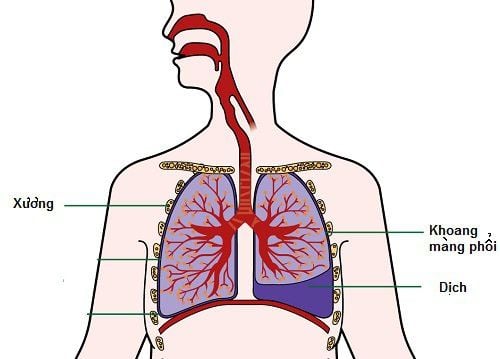
A pleural tube is a procedure in which a small tube is placed into the patient's pleural space (the space between the lungs and chest) to drain the air and fluid produced by certain diseases out of the body. body.Intubation of a pleural tube is often done to treat the following conditions:
Trauma to the chest or after chest surgery; There is air that leaves the lungs and enters the pleura and accumulates there; There is fluid accumulation in the pleural space due to chest bleeding or fluid accumulation from a chest abscess, fluid accumulation due to heart failure.
2. Characteristics of the pleural drainage tube
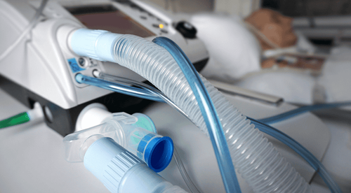
2.1 Types of pleural drainage tubes There are many types of pleural drainage tubes used such as: Gastric tubes (completely sterile), silicone pleural drainage tubes, specialized pleural drainage tubes, .. Standard drainage tubes, specifically designed for pleural drainage, have the greatest advantage because of their ease of operation, reduced complications, and reduced drainage efficiency.2.2 Size of the pleural drainage tube The size of the drain placed into the pleural space depends on each specific disease. They are:
For patients with chronic lung disease with empyema, pneumothorax or hemothorax: Usually a large pleural drainage tube with a diameter of 8-10mm is indicated; For patients with first-time pneumothorax, no chronic lung disease: Usually a pleural tube is placed with a smaller size, about 4 - 8mm in diameter. 2.3 Placement of the pleural drainage tube Normally, the pleural drain is placed in the IV intercostal space, anterior axillary line.
3. Procedure for placing a pleural drainage tube

Quy trình đặt ống dẫn lưu màng phổi như thế nào?
The doctor does a clinical examination, combined with imaging diagnostic methods such as: X-ray, chest ultrasound, chest CT scan (computerized tomography) to confirm whether there is fluid or gas accumulation. is the cause of the problem on the lungs and determine where to place the drain.
3.2 Performing the procedure A draining procedure usually takes a minimum of 20 minutes. The procedure is done as follows:
The doctor disinfects a large area on the chest: From the armpit through the nipple and close to the abdomen; The doctor spreads a sterile cloth over the disinfected area and injects anesthetic into the disinfected area; The doctor uses a scalpel to make a small incision between the 2 ribs on the chest with a length of 1-2cm, using hands and clamps to slowly separate the incision; When the incision has opened wide enough, the doctor inserts a pleural drainage tube (this is a one-way drainage tube, only sucking fluid and air from the pleura out, not allowing them to move back). that fixed stitch; Connect a continuous drain suction machine. 3.3 After the procedure The patient stays in the hospital until the drain is removed; Every day, the nurse will carefully check the drainage tube for air or fluid leaks, whether the patient has difficulty breathing; The patient is instructed to breathe deeply and cough to help expand the lungs, allowing air and fluid to escape, and preventing fluid from entering the lungs; Keep the tube to drain fluid and air always straight, not be pressed, must be placed under the lungs so that the air can escape, the lungs can expand normally. Some possible complications before, during and after pleural drainage include: Local pain, shock due to hypervagal reaction, pneumothorax, empyema and pleural bleeding. Complications of pneumothorax, severe pleural bleeding can be life-threatening.
The patient should call the doctor immediately if the following symptoms are present:
The drain is removed or displaced; The drains are broken; The patient suddenly feels short of breath or has more pain.

Người bệnh cần gọi bác sĩ nếu đột ngột cảm thấy khó thở hoặc đau nhiều
The doctor may order the patient to take another X-ray to check the effectiveness of aspiration pleural drainage.
When a pleural drainage tube is indicated, the patient needs to absolutely cooperate with the doctor to treat the disease effectively and minimize the risk of unpredictable complications.
Please dial HOTLINE for more information or register for an appointment HERE. Download MyVinmec app to make appointments faster and to manage your bookings easily.