This is an automatically translated article.
The article was professionally consulted by Specialist Doctor I Tran Cong Trinh - Radiologist - Radiology Department - Vinmec Central Park International General Hospital. The doctor has many years of experience in the field of diagnostic imaging.Magnetic resonance imaging (MRI) is a modern imaging method used in most medical examination and diagnosis procedures, including pathologies related to limb software. Accordingly, the software MRI procedure with the injection of magnetic contrast agent will clearly show the structures or organ tissues in the body.
1. What is software magnetic resonance imaging (MRI)?
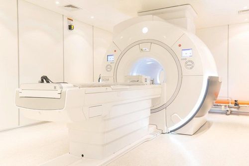
Magnetic resonance imaging (MRI) is a imaging technique that uses magnetic fields and radio waves, allowing detailed observation of lesions in terms of morphology and structure of parts in the body. The technique is reproducible, collects 3D data, and has almost no side effects. Therefore, this is one of the important tests applied in the diagnosis of diseases related to limb software.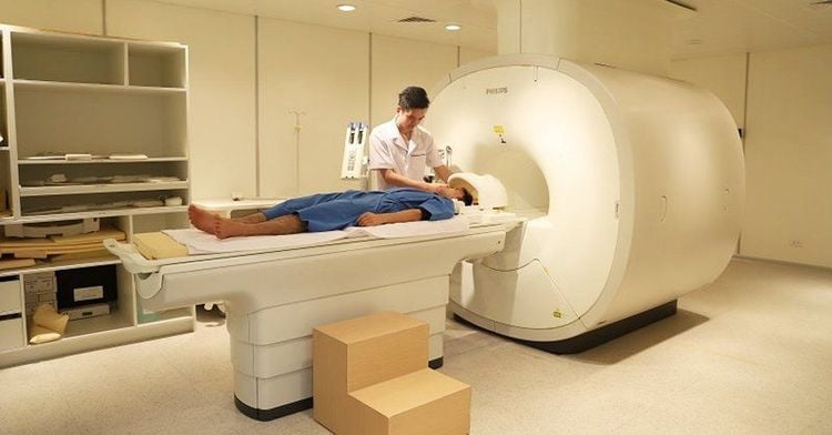
2. What are magnetic contrast drugs? What type of contrast agent is used in soft limb MRI?
Contrast is a type of contrast agent that helps to see the internal organs more clearly. Right before the MRI scan, a contrast agent is introduced into the body. The purpose is that when taken up, some images of structures or organ tissues will be displayed more clearly.Besides routes such as oral, enemas, injection into body cavities (uterus, joints, fluid spaces in the spine, etc.), intravenous contrast injection is the most commonly used. After the MRI scan for a while, the body will eliminate the contrast agent from the urinary tract and gastrointestinal tract.
Currently, the intravenous gadolinium-based contrast agent is used in the most common MRI, including soft limb MRI. When this substance enters the body, it will change the magnetic field properties of neighboring water molecules, helping to increase the contrast of internal organs, digestive tract, arteries, veins, soft tissues of the body, brain and breast.
3. What limb software pathology is indicated for MRI?
3.1 Benign tumor
Lipoma: Lipoma is a common soft tissue tumor in the extremities. Most lipomas are located in the subcutaneous tissue or between layers of muscle. When lipomas are deep, there is usually a more localized infiltrate than malignant lipomas. On MRI, the tumor is often well-defined, oval, and surrounded by a segmental margin with a thin crust. Hemangioma: Hemangioma with segmental border, low signal on T1W, very high on T2W. Hemangiomas are strongly colored after injection of magnetic contrast agent. On MRI, hemangiomas often have a normal structure, an internal vascular tuft, which may be poorly colored due to venous calcification in the hemangioma.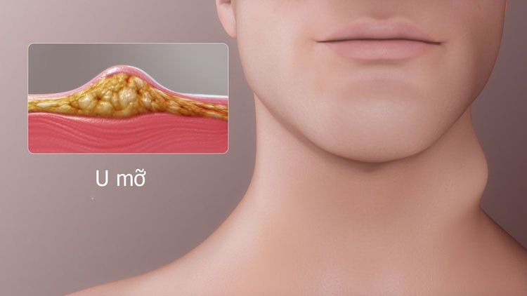
3.2 Malignant tumor
Fat sarcoma: Fatty sarcomas come in different shapes. Well-differentiated tumors are often rich in fat. In particular, malignant lipomas differ from benign lipomas in that they have many walls, soft tissue nodules inside, high signal on T2W, and strong contrast enhancement. Poorly differentiated tumors have a low fat component, making it difficult to distinguish between benign and malignant tumors. Malignant fibrous histiocytosis: Tumor located in the muscle layer, large size, unclear border, heterogeneous signal, low on T1W, high on T2W. Tumors tend to involve bone.3.3 Inflammation
For inflammation, magnetic resonance imaging helps determine the extent of involvement related to the lesion and make the differential diagnosis. Inflammatory areas often have high signal on T2W, STIR. When there is an abscess, the signal is low on T1-weighted images and high on T2-weighted images due to the presence of fluid and necrotic tissue.4. What do patients need to prepare before going for an MRI scan?
Before the injection, the patient needs to check whether he is on the list of contraindications to MRI. Contraindications to MRI include:Wearing electronic devices on the body: Pacemakers, defibrillators, ear canal implants, automatic subcutaneous injection devices. Intracranial metal surgical forceps, eye sockets and blood vessels < 6 months The patient is in poor health and requires a side resuscitation device However, the patient does not need to fast before the MRI scan. During the preparation for the scan, the doctor will thoroughly explain the procedure, change dressing instructions, and remove contraindicated items.

5. Software MRI procedure only with contrast injection
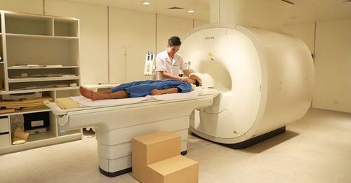
4.1 Place the patient on the MRI machine
The patient is placed in the supine position Install the receiver coil over the whole body Wear noise-canceling headphones for the patient (if necessary).4.2 Conducting an MRI scan
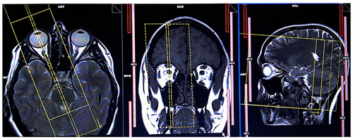
Imaging of three-plane localized pulse sequences Two-way imaging of NATIVE (Siemens) and TRACE (Philips) Multi-directional (MPR) and three-dimensional imaging (VRT) software MRI with injection Magnetic contrast is a modern and useful imaging method because it provides high-resolution images, helping doctors to diagnose the stage of the disease, thereby directing examination and treatment in a timely manner.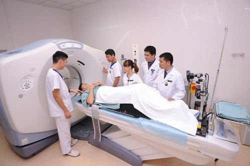
Silent technology is especially beneficial for children, the elderly, and medically ill patients. Healthy and weak patients and patients undergoing surgery Limit noise, create comfort and reduce stress for customers during the shooting process, help capture better quality images and shorten shooting time. Magnetic resonance imaging technology is the technology applied in the most popular and safest imaging method today because of its accuracy, non-invasiveness and non-X-ray use.
Please dial HOTLINE for more information or register for an appointment HERE. Download MyVinmec app to make appointments faster and to manage your bookings easily.


