This is an automatically translated article.
This article was written by Specialist Doctor II Khong Tien Dat, Doctor of Radiology - Department of Diagnostic Imaging - Vinmec Ha Long International Hospital. The doctor has a lot of experience with more than 14 years working in the field of diagnostic imaging.The main cause of pleural thickening is pleural effusion. Therefore, when identified and definitively treated, it also means stopping the pleural effusion.
1. What is pleural thickening on radiographs?
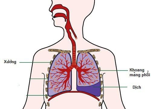
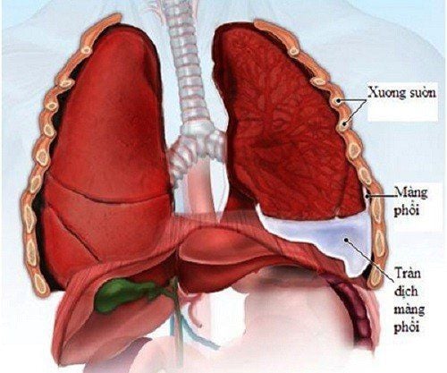
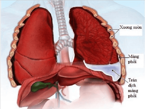
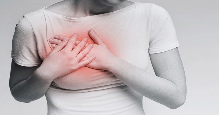
Normally, the visceral pleura and thin-walled pleura are close together, not visible on chest x-ray. When the pleural effusion is exuding, the pleural cavity is filled with fluid, the pleura is thickened, creating a clear pleural image on the film.Chest pain is the primary and typical symptom of pleural effusion. Dull pain on the side of the effusion, especially when lying on that side, the pain will increase. In addition to chest pain symptoms such as shortness of breath is also a common symptom in pleural effusion; Fever is often a symptom of an infection caused by a microorganism that causes the body to react.
2. Signs of pleural thickening on X-ray images
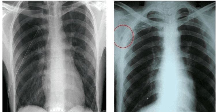
Pleural thickening on chest radiographs is often the result of pleural effusion. These signs include:Thickening of the pleura: The opacities are homogeneous or relatively homogeneous, the boundaries are often clear. Causes contraction of related organs (pharyngeal dome, mediastinum, intercostal space, ..). Thickening of the entire pleura on one side: Usually due to sequelae of poorly treated pleural effusion, hemothorax due to trauma. Solid opacity of the entire lung causes a very strong tug-of-war effect. Pulling the mediastinum toward the lesion, the intercostal space on the injured side is narrowed. There may be calcification of the pleural space.
Trắc nghiệm: Làm thế nào để có một lá phổi khỏe mạnh?
Để nhận biết phổi của bạn có thật sự khỏe mạnh hay không và làm cách nào để có một lá phổi khỏe mạnh, bạn có thể thực hiện bài trắc nghiệm sau đây.3. Treatment of thickened pleural fluid
Pleural thickening is mainly caused by pleural effusion. Therefore, when identified and definitively treated, it also means stopping the pleural effusion.Pleural effusion is usually treated by aspiration pleural aspiration to do tests and help the patient breathe easier. Once the cause has been identified, it is essential to treat the underlying cause to reduce or stop the pleural effusion.
After treatment of effusion, it is necessary to intervene with anti-pleural drugs because the most common consequence of pleural effusion is to cause thickening and adhesion of the pleura, which greatly affects respiratory function.
The early detection of the disease makes the treatment highly effective, so when there are signs such as shortness of breath, increased chest pain, etc., the patient should not be subjective but need to go to a medical facility to diagnose the disease early. .
Please dial HOTLINE for more information or register for an appointment HERE. Download MyVinmec app to make appointments faster and to manage your bookings easily.


