This is an automatically translated article.
The article is professionally consulted by Master, Doctor Le Anh Viet - Radiologist - Department of Diagnostic Imaging and Nuclear Medicine - Vinmec Times City International Hospital.Spectroscopy is an important technique in the diagnosis and treatment of dementia in particular and many neurological diseases in general.
1. Dementia overview
Dementia is a neurodegenerative disease that causes a decline in cognitive function of the brain, from which cognitive disorders, behavioral and language disorders... affect the quality of life, even activities. patient's self-care. Dementia usually occurs in the elderly, over 60 years old, but now also occurs more in young people.2. Dementia symptoms
Symptoms of dementia at the beginning are often difficult to recognize, patients only feel signs such as:● Forgetful, memory loss, or lost.
Having difficulty in communication.
● Daily activities are slow and ineffective.
● Inability to learn or remember new information.
Having difficulty with planning and organization.
● Lack of ability to proactively solve unexpected problems.
Difficulty coordinating with others.
● Change in personality (anxiety, hallucinations, paranoia...)

Sa sút trí tuệ gây lên các dấu hiệu như hay quên, mất trí nhớ ở người mắc phải
3. Causes of dementia
Increasing life expectancy, increasing rates of many chronic diseases are the factors driving the increasing number of elderly people with dementia. There are also many causes of dementia such as:Diseases with many potential risks of blood vessels (high blood pressure, diabetes, cardiovascular disease, cerebrovascular accident, ..).
● Hypothyroidism (vitamin B deficiency, nutritional deficiency).
● Long-term alcohol abuse leads to poisoning, atherosclerosis complications, brain atrophy and dementia.
● Less mental and physical activity often.
Genetic factors.
Alzheimer's disease, Huntington's disease, Down's disease.
Tumors in the brain.
4. Diagnosing Dementia
Depending on the case, the doctor will order additional tests to make an accurate diagnosis:● Clinical examination.
● Ask the patient to perform a neuropsychological test.
● Assess the nervous system by CT tomography, magnetic resonance imaging (MRI), cranial magnetic resonance spectroscopy.
EEG EEG .
Analysis of cerebrospinal fluid.
Blood and urine tests...
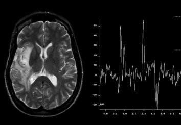
Chụp cộng hưởng từ phổ não giúp các bác sĩ chẩn đoán sa sút trí tuệ
5. What is magnetic resonance brain spectroscopy?
Cranial magnetic resonance imaging is a new technique used in the diagnosis of neuropathies, helping to detect specific metabolites of traumatic brain injury. Cranial magnetic resonance imaging is based on the principle of comparing the concentrations of metabolites in the examination area to determine the nature of the lesion.6. Brain spectrum magnetic resonance imaging procedure
6.1 Preparation before the procedure To perform cranial magnetic resonance imaging, it is necessary to prepare:Implementation team:
● Radiology specialist.
Electro-optical technician.
● Nursing.
Vehicles used:
Resonance angiography machine from 1 Tesla or more.
Film printer.
Movies.
Image storage system.
Drugs: prepare sedatives.
Patients need to prepare:
● No need to fast.
● Prepare a clinician's scan order with a clear diagnosis or complete medical record (if needed).
● The procedure is explained in advance to coordinate well with the doctor.
● Check for contraindications.
● Change the clothes of the MRI room according to the instructions, remove the contraindicated items.
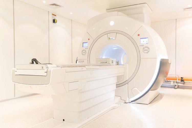
Hệ thống máy chụp cộng hưởng từ 1 Tesla trở lên trong chụp cộng hưởng từ phổ não
Patient position
● The patient is placed supine on the imaging table.
● Move the shooting table into the machine bay.
Capture technique
Locate the measuring area.
● Select pulse sequences suitable for examination purposes.
● Perform normal pulse sequences: T1, T2, Flair on all subjects.
● Selected cutting directions: horizontal (horizontal), horizontal (horizontal) and vertical (vertical).
Note: Other special pulse sequences may be performed to diagnose specific brain conditions (if needed). For example, the T2* pulse sequence helps identify bleeding in the lesion.
● Intravenous contrast is injected, and T1 pulses are taken in 3 directions without dilatation to evaluate the enhancement of the lesion (central area to be examined).
● Place the measuring cell ( multi voxel or mono voxel ) in the area to be measured. Set TE time suitable for each purpose, set anti-interference frame.
Note: Avoid the area close to the bone, close to the bleeding area. If taking multi voxel, the imaging area will include the lesion, surrounding edema and the opposite healthy area to compare the difference in metabolite concentrations.
● Proceed to pulse and process the obtained images, select the necessary images showing the pathology for film printing or disc burning.
● Based on the received image, the doctor reads the lesion and evaluates the results.
The imaging results of cranial magnetic resonance imaging are often analyzed as a histogram, characterized by information about frequency, height, width, area, height, or area below the peak of the body. show concentrations of metabolites. Common metabolites commonly imaged on the spectrum are Choline, Creatine, NAA and Lactate. Multiple scans can help monitor changes in metabolite levels, leading to more accurate diagnostic conclusions.

Người bệnh được đặt nằm ngửa trên bàn chụp trước khi tiến hành chụp cộng hưởng từ phổ não
7. Role of brain spectroscopy in the diagnosis of dementia.
The acquired brain spectrum imaging plays an important role in the evaluation of patients with suspected dementia. The earlier the lesions are detected, the more effective the treatment. When performing the technique, the patient should follow the instructions of the doctor to achieve high diagnostic efficiency and provide appropriate treatment.Vinmec International General Hospital is a high-quality medical facility in Vietnam with a team of highly qualified medical professionals, well-trained, domestic and foreign, and experienced.
A system of modern and advanced medical equipment, possessing many of the best machines in the world, helping to detect many difficult and dangerous diseases in a short time, supporting the diagnosis and treatment of doctors the most effective. The hospital space is designed according to 5-star hotel standards, giving patients comfort, friendliness and peace of mind.
Master Doctor Le Anh Viet has 07 years of experience working in the field of diagnostic imaging, performing well in diagnostic imaging techniques. Currently, the doctor is working at the Department of Diagnostic Imaging and Nuclear Medicine - Vinmec Times City International General Hospital.
To register for an examination at Vinmec International General Hospital, you can contact the nationwide Vinmec Health System Hotline, or register online HERE.


