This is an automatically translated article.
Posted by Master, Doctor Mai Vien Phuong - Department of Examination & Internal Medicine - Vinmec Central Park International General Hospital
Increasing evidence has demonstrated a role for RON in regulating cancer cell invasion, which is related to the effects of RON activation on a wide range of signaling mechanisms.
1. Evidence has demonstrated a role for RON in regulating cancer cell invasion
Activation of complex downstream signaling networks including signal transducers and transcriptional activators, β-catenin, JUN N-terminal kinase, MAPK, and PI3K/AKT pathways are major contributors part of the RON-mediated aggressive cancer phenotype. In breast cancer, several signaling pathways important for stem growth, invasion, and proliferation are activated by RON. For example, the cytoplasmic antigenic antigen (PCNA) pathway of RON-cellular Abelson murine leukemia cells was identified to contribute to RON-mediated cell proliferation in breast cancer.Unregulated RON signaling leads to c-Abl activation, which in turn leads to PCNA phosphorylation. Furthermore, in breast cancer, RON signaling regulates cancer cell invasion through the activation of DEK proto-oncogene (DEK), a DNA-binding non-histone nuclear phosphoprotein that induces closed circular DNA closed to form positive superhelixes. This process appears to work through a canonical and endocrine β-catenin signaling loop that ultimately influences breast cancer angiogenesis. In addition, RON-dependent PI3K upregulation of the methyl-CpG 4 binding region, DNA glycosylase (MBD4) increases invasive growth and metastasis of breast cancer cells through methylation reprogramming. DNA of specific target genes. Clinical data indicate that in breast cancer patients, poor prognosis is associated with the RON-MBD4
epigenetic pathway.
2. RON receptor and hepatobiliary carcinoma
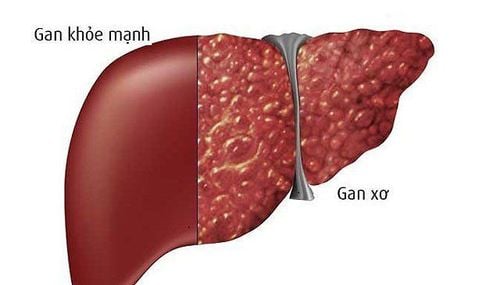
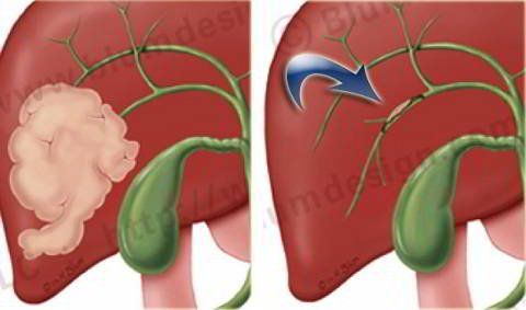
By 2030, it is estimated that more than 1 million people will die from liver cancer worldwide. Major malignancies of the liver and adjacent biliary tract include HCC, intrahepatic and extrahepatic cholangiocarcinoma (CCA), and gallbladder cancer (GBC). Among them, HCC and CCA in the liver accounted for 85% and 10%, respectively. Abnormal RON expression was observed in HCC, possibly related to the pathological status of this cancer. In an HCC cell line study, RON was shown to be associated with oncogenic and invasive phenotypes (eg, resistance to apoptosis, cell migration) tumor cells and tumor cell invasion) through the modulation of the AKT signaling cascade, c-Raf, and extracellular regulatory kinase (ERK). Clinically, RON and MET expression in HCC patients after surgical resection showed no association of RON with overall survival and recurrence. However, patients with RON+/MET+ disease had a higher overall recurrence rate than patients with alternate presentation.

Trong nghiên cứa ung thư biểu mô tế bào gan và thụ thể RON có liên quan đến các kiểu hình gây ung thư và xâm lấn
3. RON receptor is emerging as an important mediator of CCA cholangiocarcinoma pathogenesis and clinical prognosis
Similar to HCC, RON is emerging as an important mediator of CCA pathogenesis and clinical prognosis. Investigation of RON and MET expression in perioral CCA patients who underwent histological resection revealed that patients with RON+/MET+ disease showed poorer overall survival than patients with other samples. In addition, in patients with extrahepatic CCA, complete loss of MET, RON, or both (and their overexpression) is a poor prognostic factor, possibly due to the high rate of lymph node metastasis.Recently, Cheng and colleagues showed that BMS-777607, a dual inhibitor of MET-RON, inhibits human CCA cell growth HuCCT1 and KKU-100, and reduces tumor growth. tumors in CCA mice. They also found that for patients with CCA who had previously undergone hepatectomy, adjustment for RON and MET was a poor predictor of survival. Taken together, these studies suggest that the aberrant RON expression found in human hepatobiliary carcinoma cell lines and samples is strongly associated with pathological status and clinical outcome.
4. RON receptor and pancreatic cancer
The majority of malignancies of the pancreas are adenocarcinomas, of which pancreatic ductal carcinoma is the most common malignancy, accounting for more than 95% of all malignancies of the pancreas . Pancreatic cancer poses a serious health problem and has an extremely poor prognosis because of its patient-specific symptoms, its strong and substantial resistance to most conventional treatments. often, and the fact that it has many genetic and epigenetic variations. Therefore, there is a need for new therapies to treat pancreatic cancer. In recent years, the function of RON in pancreatic cancer has been widely defined in many model systems, such as animal, cellular, and clinical bases.
To date, researchers have reported that RON is expressed in different pancreatic cancer cell lines, such as CFPAC-1, ASPC-1, Hs766.T, L3.6pl, HPAFII, HPAC, Capan-2 and BXPC-3. However, MIA-PACA-2 cells showed minimal RON expression. The association of RON with Kras-driven pancreatic carcinogenesis was investigated using transgenic mouse models. The results showed that overexpression of RON accelerates pancreatic epithelial neoplasia (PanIN) progression, enhances tubular metaplasia, and promotes tumor progression for invasive pancreatic cancer. encroachment. Furthermore, the study demonstrated that initiation of PanIN was slowed by inactivation of the RON kinase gene, leading to smaller tumors and ultimately prolonging survival of tumor-bearing mice.

Thụ thể RON sẽ thúc đẩy sự tiến triển của khối u đối với bệnh nhân ung thư tuyến tụy xâm lấn
5. Advances in understanding the clinical relevance of RON in pancreatic cancer
The authors have made great progress in the understanding of the clinical relevance of RON in pancreatic cancer, mainly focusing on the status of RON expression in pancreatic cancer samples and its potential uses. its potential as a prognostic biomarker for patient survival. IHC staining using anti-RON antibodies is a commonly used approach to evaluate RON expression in different experimental settings. Several studies have identified positive sample rates in pancreatic cancer specimens as 70%, 88%, 96%, 80% and 86%, respectively. Meanwhile, in pancreatic cancer samples, high RON expression was detected, while minimal levels were detected in their respective normal epithelial cells.
6. During the progression of pancreatic adenocarcinoma, the frequency and level of RON expression increases
Among human pancreatic tissue samples, RON expression was detected in 83% of metastatic lesions, 79% of primary lesions and 93% of high-grade PanINs using immunohistochemistry, with expression Low levels were detected in low-grade and normal PanIN ducts (18% and 6%, respectively), suggesting that RON may be active in pancreatic carcinogenesis and metastatic progression. Furthermore, RON expression levels were significantly associated with overall survival in pancreatic cancer patients, suggesting that RON may be an important indicator for pancreatic cancer prognosis. Conflicting results between RON expression and pancreatic cancer prognosis were found in an initial study, therefore further research is needed to determine the utility of RON as a prognostic biomarker in patients. Pancreatic Cancer.
Please dial HOTLINE for more information or register for an appointment HERE. Download MyVinmec app to make appointments faster and to manage your bookings easily.


