This is an automatically translated article.
The article was professionally consulted by Specialist Doctor I Nguyen Thanh Hai - Radiologist - Department of Diagnostic Imaging and Nuclear Medicine - Vinmec Times City International General Hospital.Prostate magnetic resonance imaging is a procedure applied in the diagnosis and evaluation of prostate lesions to detect disease and take timely and effective treatment measures for patients.
1. Function of the prostate
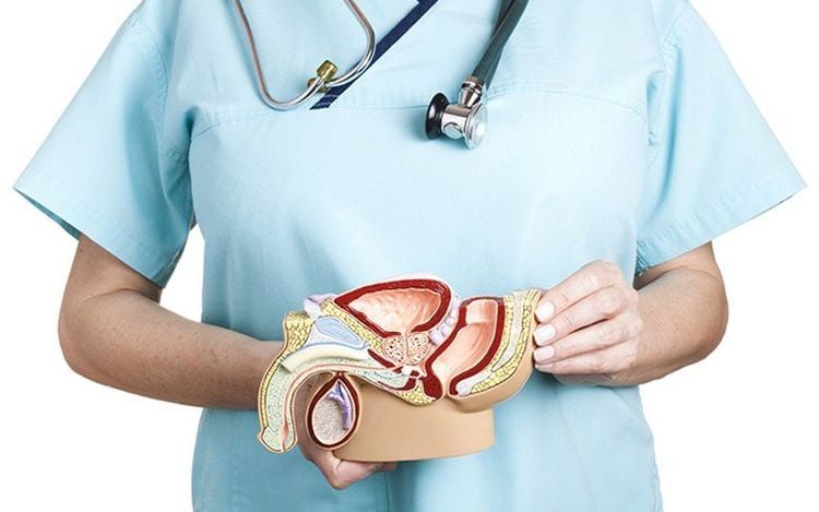
The prostate gland is an organ located below the bladder, wrapped around the tip of the urethra just below the bladder. The prostate gland in an adult male weighs about 15-25g, averages about 4cm wide, 2.5cm thick and 3cm high.The prostate gland is composed of 70% glandular tissue and 30% fibromuscular stroma. The cushion that surrounds the prostate gland, contracts during ejaculation, and pours secretions from the prostate into the urethra.
In boys, the prostate gland is as small as a pea and does not take on any tasks for the body. During adolescence and young adulthood, the prostate gland grows stronger. The prostate gland's job is to secrete fluid to form semen, and at the same time provide energy for sperm so that sperm can fertilize an egg. In addition, the prostate gland also plays an important role in urinary control in men.
Common prostate diseases: inflammation, prostate enlargement, prostate cancer.
2. Detailed procedure for magnetic resonance imaging of prostate gland
Prostate magnetic resonance imaging helps doctors assess the concentration of metabolites in localized prostate lesions, contributing more information to help doctors distinguish prostate enlargement early. benign, focal inflammatory lesions or prostate cancer.2.1 Indications ● Prostate cancer suspected cases after other imaging and testing methods have been performed.
● Perform prostate MRI at the professional request of the treating doctor.
2.2 Contraindications Absolute contraindication (general contraindication for magnetic resonance imaging).
● People with serious illnesses need resuscitation equipment on their side;
● People with metal surgical clips in the eye socket, intracranial or vascular for less than 6 months;
● People wearing electronic devices such as pacemakers, anti-vibration machines, automatic subcutaneous drug injection devices, cochlear implantation,...
Relative contraindications
● Persons with metal surgical forceps for more than 6 months;
People who are afraid of the dark or afraid of being alone.
2.3 Preparation before MRI ● Personnel: Including specialists, radiology technicians and nurses;
● Drugs: Including contrast agents, sedatives and skin and mucosal antiseptics;
● Medical supplies: 10ml syringe, 18G intravenous needle, physiological saline or distilled water, cotton, gauze, sterile gloves and bandages, medicine box, emergency equipment for drug accidents optical ;
● Vehicles: Resonance angiography machine of 1 Tesla or more; film, film printers and image storage systems;
● Patient: Technical explanation is given; checked for contraindications; no need to fast; change the clothes of the magnetic resonance imaging room, remove contraindicated items as prescribed; Bring your doctor's request for an MRI of the prostate or complete medical records if needed.
2.4 Carrying out the technique
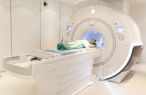
● Routine imaging technique:
Place an intravenous line with an 18G needle, connected to a double-bore electric injection pump (1 barrel contains physiological saline, 1 barrel contains magnetic contrast). The dose of contrast agent used is 0.1mmol/kg body weight;
Capture positioning;
○ Take before injecting magnetic contrast agent: Take pulse sequence 1 - 2 - 3 - 4 according to technical requirements;
○ Take a picture after injecting magnetic contrast: Perform contrast injection with dose 0.1mmol/kg body weight, speed 2ml/sec. Next, capture the 5th - 6th pulse sequence according to the specifications;
● Spectral Magnetic Resonance Technique:
Spectral Magnetic Resonance Pulse can be single-volume or multi-volume;
○ Determine the concentration of metabolites including Lipid, Choline, Lactate,...;
○ The technician prints the film and transfers the images to the doctor;
○ The doctor analyzes the images and parameters of the magnetic resonance spectrum to make diagnostic conclusions.
Note: The rectal signal coil or the abdominal wall receiver coil (in the case of thin patients) can be used on the abdomen in the pelvic region, fixed with a lifeline and using a small field of view.
2.5 Evaluation of results ● On magnetic resonance imaging, the entire prostate gland and nearby organs such as rectum, seminal vesicles,...;
● Determination of metabolite concentrations and ratio between metabolites;
● Doctors read injuries and give professional advice to patients and relatives.
2.6 Some complications and how to deal with them ● The patient may be agitated or scared, the doctor should encourage and comfort the patient;
● In case the patient is too worried or afraid, the doctor can give the patient a sedative under the supervision of the anesthesiologist;
● With adverse events related to contrast agents such as anaphylaxis, nausea, hypotension, urticaria, laryngeal edema,... need to be handled according to the standard protocol.
When performing magnetic resonance imaging of the prostate, the patient should follow all instructions given by the physician. This helps to diagnose quickly, effectively and reduce possible risks.
Vinmec International General Hospital is a high-quality medical facility in Vietnam with a team of highly qualified medical professionals, well-trained, domestic and foreign, and experienced.
A system of modern and advanced medical equipment, possessing many of the best machines in the world, helping to detect many difficult and dangerous diseases in a short time, supporting the diagnosis and treatment of doctors the most effective. The hospital space is designed according to 5-star hotel standards, giving patients comfort, friendliness and peace of mind.
Doctor Hai has more than 20 years of experience in the field of diagnostic imaging, especially in the field of multi-segment computed tomography, magnetic resonance. Currently, the doctor is working at the Department of Diagnostic Imaging and Nuclear Medicine - Vinmec Times City International General Hospital.
Please dial HOTLINE for more information or register for an appointment HERE. Download MyVinmec app to make appointments faster and to manage your bookings easily.


