This is an automatically translated article.
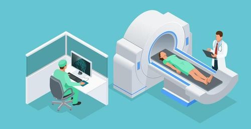
This article is professionally consulted by Master, Resident Doctor Tran Duc Tuan - Department of Diagnostic Imaging - Vinmec Central Park International General Hospital.Thoracic resonance imaging with injection of magnetic contrast is one of the diagnostic methods with high value in diseases of the lung, mediastinum and chest wall. In lesions where investigation of the lesion is required and blood supply is assessed, contrast agents can help define the requirements for investigation of vascular distribution and tissue perfusion. So what is the procedure for chest magnetic resonance imaging with magnetic contrast injection?
1. Outline
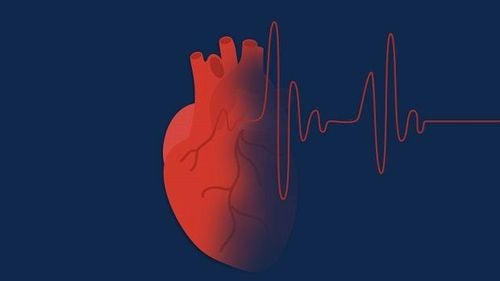
Thoracic anatomy is divided into two main parts including the thoracic cavity containing the essential organs of the body and the chest wall consisting of the layer of muscle, bone and skin that protects the organs in the chest cavity. The cardiovascular system includes the heart and large blood vessels such as the aortic arch, the ascending and descending vena cava, and the dissecting arterioles located in the mediastinum. In addition, the organs of the respiratory system including the lungs and airways are located in the chest cavity. Both are vital organs of the body. There are also lymphatic and spinal nerve systems in the back.
Chest magnetic resonance imaging with injection of magnetic contrast helps to increase the contrast of blood vessels and the doctor can assess the level of tissue perfusion through the time of contrast enhancement (from the arteries to the capillaries). vessels in the tissue examined and returned to the vein). Therefore, diseases related to the thorax need to assess the location, size, density and blood supply level, and magnetic resonance imaging with contrast injection is very valuable in diagnosis and treatment prognosis.
2. Designation
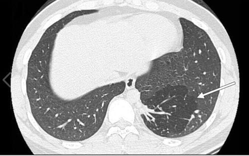
Survey the characteristics, location, size, density of pathological lesions in the lung, mediastinum, chest wall; Evaluation and survey of blood vessels in the chest area, the extent of proliferation of the lesions; Some indications depend on the clinical picture and the specific requirements of the treatment.

Bác sĩ điều trị sẽ là người đưa ra chỉ định chụp MRI phù hợp với tình trạng bệnh lý của bệnh nhân
3. Contraindications
3.1. Absolute contraindications Absolute contraindications of magnetic resonance imaging: implantation of metal-based electronic devices such as pacemakers, anti-vibration machines, cochlear electrodes, automatic continuous injection machines under skin, implantation of means to fix bones with metal intracranial, orbital, vascular < 6 months. Metal foreign body in chest area. Severe advanced disease requires emergency resuscitation equipment for monitoring and treatment. Absolute contraindications related to the contrast agent: allergy, hypersensitivity or anaphylaxis to the contrast agent. 3.2. Relative contraindications Metal surgical facilities > 6 months; Fear of the dark, fear of narrow spaces, fear of loneliness; Renal failure with glomerular filtration rate <30 mL/min, pregnant or lactating women.
4. Procedure of chest magnetic resonance imaging with injection of magnetic contrast agent
4.1 Preparation before proceeding Performer: radiologist and radiology technician. Equipment: 1.5 Tesla or larger resonator Prepare disinfectant, syringe and magnetic contrast agent. Drugs: Sedative support drugs can be used in cases of patients with fear of the dark, claustrophobia of narrow spaces. Patient: can have a light meal, it is not necessary to fast before the scan. Instructions and explanations of what to do before filming begins. Patient removes contraindicated items.

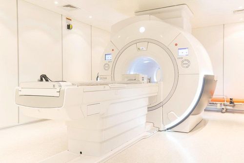
Hệ thống máy chụp cộng hưởng 1.5 Tesla
4.2. Steps to conduct chest magnetic resonance imaging with magnetic contrast injection 4.2.1 Patient's position
Lying supine on the magnetic resonance imaging table; Position the signal receiver; Adjust the table to the sewing compartment and locate the chest area to be investigated; Adjust the controller to capture breathing, reduce image noise; In case the lesion is located in the chest wall, the position can be adjusted, placing the patient on the side of the lesion to avoid infection due to breathing. 4.2.2. Sequences of pulses before contrast injection from
Initial localization capture the entire thorax (starting from 1st cervical vertebra to the end of the diaphragm in all three planes); T2W pulse series of horizontal and transverse sections (from the apex of the lung to the angle of the costal ribs), thickness of each slice from 6 to 8 mm, interval 10-20% of the cut layer, FOV 380-400, can use magnetic shield if necessary, phase difference from LR (left – right); The T1W pulse sequence, the cross-sectional plane is similar to the cross-sectional T2W pulse sequence. 4.2.2. Pulse sequences after injection of magnetic contrast
T1W pulse sequence, cross-sectional and horizontal planes after injection of magnetic contrast agent (dose 0.1 mmol/kg body weight at injection rate of 2 mL/s), note the need to taken immediately after injection.
4.3 Reading results Magnetic resonance imaging must meet the criteria to clearly visualize chest anatomical structures on selected pulse sequences; Detect the location, size, density of the lesion and assess the extent of contrast enhancement in the tissue to be investigated; The doctor specializing in diagnostic imaging is the person who reads the lesions and describes them on the computer with the photographic film stored; Print movies, save CDs (if needed) and diagnostic results; Get expert advice on results and recommend further testing if needed.

Phim chụp cộng hưởng cần được bác sĩ chuyên khoa chẩn đoán hình ảnh đọc kết quả
5. Follow-up during and after magnetic resonance imaging
During magnetic resonance imaging: closely monitor vital signs (pulse, blood pressure, breathing rate), consciousness and focal neurological signs of the patient. After the magnetic resonance imaging process: the patient may be allowed to lie down for 15 minutes to observe if there are abnormalities during the scan (the patient is excited, the vital signs change) or transfer to the emergency unit for further monitoring.
6. Complications and treatment directions
Some patients with agoraphobia, solitudeism, or claustrophobia may be fearful and agitated. The technician or doctor needs to guide and encourage the patient to comfort or use sedatives in case of indication. For adverse events related to contrast agents, it is important to recognize them early to diagnose and manage contrast injection complications. Vinmec International General Hospital is currently one of the major hospitals with modern machinery and equipment for general medical examination and treatment and accurate and modern MRI for brain and vascular diseases. brain in particular.
Especially, Vinmec International General Hospital is the first unit in Southeast Asia to put into use the new 3.0 Tesla Silent Resonance Imaging machine from the US manufacturer GE Healthcare.
The machine currently applies the safest and most accurate magnetic resonance imaging technology available today, without using X-rays, non-invasive. Silent technology is very beneficial for patients who are young children, the elderly, patients with weak health or have just had surgery.
Master, Doctor Tran Duc Tuan is well-trained in the country and many centers have prestigious medical background in the world such as: Australia, Singapore, Thailand... The doctor has many years of experience in the field of medicine. imaging and endovascular and extravascular interventions. Currently, the doctor is working at the Department of Diagnostic Imaging and Interventional Vascular Unit - Vinmec Central Park International General Hospital.
To register for an examination at Vinmec International General Hospital, you can contact the nationwide Vinmec Health System Hotline, or register online HERE.