This is an automatically translated article.
This article is professionally consulted by Master, Resident Doctor Tran Duc Tuan - Department of Diagnostic Imaging - Vinmec Central Park International General Hospital.The thorax is an important structure of the body with many essential organs such as the heart, lungs, large blood vessels, mediastinum. Magnetic resonance imaging of the chest without contrast injection can help determine the location, extent, and size of organ lesions within the thorax. So what is the procedure for chest magnetic resonance imaging without injecting magnetic contrast?
1. Outline
The chest is an anatomical part containing many important organs, separated from the abdomen by the diaphragm, including two main components, the chest wall and the thoracic cavity.
The thoracic cavity contains the heart and a system of large blood vessels such as the aorta, vena cava, pulmonary arterioles and lymphatic system located in the mediastinum. In addition, the chest cavity also contains the organs of the respiratory system such as lungs, trachea, diaphragm. In front of the heart is a thymus gland that is involved in the body's immune system.
Magnetic resonance imaging has high value in surveying soft tissues, skeletal muscles in the chest wall and organs located deep in the mediastinum and lungs. Therefore, in the pathologies related to the anatomical structure of the thorax, magnetic resonance imaging without injection has high value in diagnosis and treatment prognosis for patients.
2. Designation
Survey the characteristics, location, size, density of pathological lesions in the lung, mediastinum, chest wall. Some indications depend on the clinical picture and the specific requirements of the treatment.

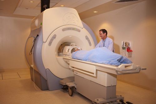
Bệnh nhân có bệnh lý phổi cần được chỉ định chụp MRI lồng ngực
3. Contraindications
3.1. Absolute contraindications Implant electronic devices with metal such as pacemakers, anti-vibration machines, cochlear electrodes, automatic continuous subcutaneous injection machines; Implantation of means of bone fixation with metal intracranial, orbital, vascular < 6 months; Metal foreign body in chest area; Severe progression of the disease requires emergency resuscitation equipment for monitoring and treatment. 3.2. Relative contraindications Metal surgical facilities > 6 months; Fear of the dark, fear of narrow spaces, fear of loneliness.
4. Procedure of thoracic resonance imaging without injection of magnetic contrast agent
4.1 Preparation before proceeding Performer: radiologist and radiology technician. Equipment: resonator 1.5 Tesla or larger Medicine: can be used to support sedation in cases of patients with fear of the dark, claustrophobia. Patient: It is not necessary to fast before the scan. Instructions and explanations of what to do before filming begins. The patient removes items that are contraindicated for magnetic resonance imaging.

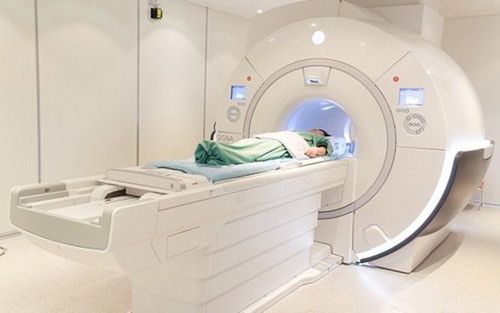
Hệ thống máy chụp cộng hưởng từ 1.5 Tesla
4.2. Steps to conduct magnetic resonance imaging of the thorax without injection of magnetic contrast agent 4.2.1 Patient's position
Lying supine on the magnetic resonance imaging table; Position the signal receiver; Adjust the table to the machine compartment and locate the chest area to be examined; Adjust the controller to capture breathing, reduce image noise; In case the lesion is located in the chest wall, the position can be adjusted, placing the patient on the side of the lesion to avoid infection due to breathing. 4.2.2. Selected pulse sequences
Initial positioning capture of the entire thorax (starting from the 1st cervical vertebra to the end of the diaphragm in all three planes); T2W pulse series of horizontal and transverse sections (from the apex of the lung to the angle of the costal ribs), thickness of each slice from 6 to 8 mm, interval 10-20% of the cut layer, FOV 380-400, can use magnetic shield if necessary, phase difference from LR (left – right); The T1W pulse sequence, the cross-sectional plane is similar to the cross-sectional T2W pulse sequence. 4.3 Reading results Magnetic resonance imaging must meet the criteria to clearly visualize chest anatomical structures on selected pulse sequences; Detect location, size, density of lesions; The doctor specializing in diagnostic imaging is the person who reads the lesions and describes them on the computer with the photographic film stored; Print film and diagnostic results; Get expert advice on results and recommend further testing if needed.

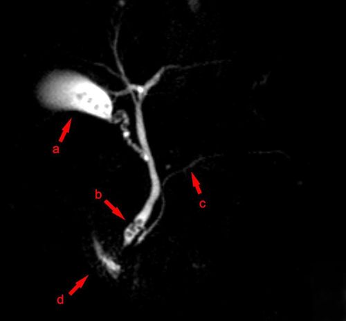
Kết quả chụp cộng hưởng từ vùng lồng ngực không tiêm thuốc đối quang từ
5. Follow-up during and after magnetic resonance imaging
During chest magnetic resonance imaging: closely monitor vital signs (pulse, blood pressure, breathing rate), consciousness and focal neurological signs of the patient. After the magnetic resonance imaging process: the patient can be rested for 15 minutes and monitored if there are abnormalities during the scan or transferred to the emergency unit for further monitoring.
6. Complications and treatment directions
Some patients with agoraphobia, solitudeism, or claustrophobia may be fearful and agitated. The technician or doctor needs to guide and encourage the patient to comfort or use sedatives in case of indication.
Vinmec International General Hospital is currently one of the major hospitals with modern machinery and equipment for general medical examination and treatment procedures and accurate and modern MRI for brain and vascular diseases. brain blood in particular.
Especially, Vinmec International General Hospital is the first unit in Southeast Asia to put into use the new 3.0 Tesla Silent Resonance Imaging machine from the US manufacturer GE Healthcare.
The machine currently applies the safest and most accurate magnetic resonance imaging technology available today, without using X-rays, non-invasive. Silent technology is very beneficial for patients who are young children, the elderly, patients with weak health or have just had surgery.
Master, Doctor Tran Duc Tuan is well-trained in the country and many centers have prestigious medical background in the world such as: Australia, Singapore, Thailand... The doctor has many years of experience in the field of medicine. imaging and endovascular and extravascular interventions. Currently, the doctor is working at the Department of Diagnostic Imaging and Interventional Vascular Unit - Vinmec Central Park International General Hospital.
To register for an examination at Vinmec International General Hospital, you can contact the nationwide Vinmec Health System Hotline, or register online HERE.