This is an automatically translated article.
This article is professionally consulted by Dr., Dr. Tran Nhu Tu - Dean and Master, Doctor Lam Thi Kim Chi - Department of Diagnostic Imaging - Vinmec Danang International HospitalThoracic resonance imaging is a technique of imaging the chest with a scanner to diagnose diseases of the lungs, pleura, mediastinum, blood vessels, heart, and chest wall to help doctors understand the results and give recommendations. best method of treatment.
1. Chest magnetic resonance imaging
Magnetic resonance imaging is an imaging technique that uses radio waves and a magnetic field to image structural details inside the body. Magnetic resonance does not use electromagnetic radiation like in X-rays or CT scans, so it is completely harmless to the body. Accordingly, magnetic resonance imaging gives high-quality images with good resolution and contrast, helping doctors to evaluate body parts in detail and diagnose disease more accurately than other techniques. other imaging such as X-rays, ultrasounds, and CT scans.
Thoracic resonance imaging is a technique to record images of the chest to diagnose a number of lung, thoracic, and cardiac diseases... If in the past, it was relatively difficult to take pictures to evaluate the chest and chest. difficult because breathing and heart are always moving, now, patients only need to hold their breath for a short period of time, the MRI machine will quickly capture images with detailed and sharp quality, helping doctors The doctor determines the best course of treatment for the patient. Therefore, the introduction of MRI scanners has brought many benefits to patients, such as:
Not affected by radiation and biological effects Obtain multi-plane images: vertical, tilted, or horizontal Displays sharp images High resolution soft tissue Non-invasive imaging techniques Minimal side effects from contrast Vascular examination without the use of contrast

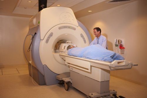
Chụp cộng hưởng từ rất an toàn cho sức khỏe
2. Indications and contraindications for thoracic resonance imaging
As a modern diagnostic method, bringing many benefits, but this method will also be indicated and contraindicated by doctors in the following cases:
2.1 Indications for chest magnetic resonance imaging Some Indications for thoracic resonance imaging include:
Suspected lesions of the chest wall, heart, lungs, mediastinum... Diagnosis of thoracic tumors Accurately assess the development stage of tumor Evaluation of aortic aneurysm , flow Evaluation of mediastinal lymph node mass Suspected damage to mediastinal chest wall, lung, heart 2.2 Contraindications to thoracic resonance imaging People carrying electronic devices or metal is present. Indispensable patient has resuscitation on the side Using metal surgical forceps Afraid of the dark, incubators, solitude in need of encouragement or sedation.
3. Steps to conduct thoracic resonance imaging
Prepare the patient's position
The patient can lie on his back on the table Select and position the receiver Move the table into the machine compartment and position the imaging area Adjust the controller to capture breathing, reduce noise image, if the lesion is in the chest wall, the patient may lean to the injured side to avoid noise due to breathing. Techniques to perform
Positioning capture the entire ribcage from the base of the neck to the end of the diaphragm in 3 directions. Transverse and horizontal T2W pulse sequence: from the apex of the lung to the angle of the costodiaphragm, section 6 -8mm, distance 10 -20% of the cut, FOV 380 -400, magnetic shielding if needed, phase difference from LR ( left and right). Cross-sectional T1W pulse sequence capture is similar to cross-sectional T2W. Print film or transfer photos to the doctor's workstation. The doctor analyzes the images and makes a diagnosis. Magnetic resonance imaging of the chest will clearly see the anatomical structures of the ribcage and organs in the chest, and detect lesions and lung diseases if any.
If the patient is excited or nervous when taking pictures, the doctors and technicians need to encourage and comfort the patient or maybe give sedation with the supervision of the anesthesiologist.
Different from the concept of using only X-ray and CT scanner for chest examinations, thoracic magnetic resonance imaging method is increasingly widely used in thoracic examination to diagnose lung and chest wall diseases. , mediastinum, blood vessels for diagnosis and timely intervention measures.
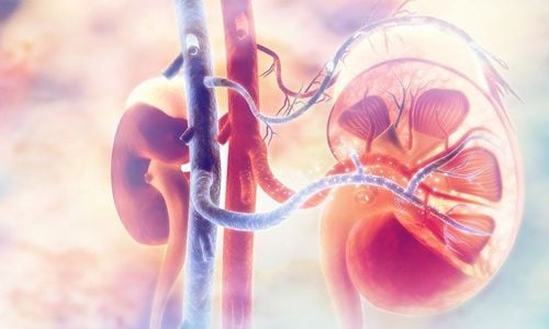
Lungs are one of the most important human organs, so early examination will detect diseases (if any), since then the treatment methods will also become gentler and simpler than in the early stages. late segment.
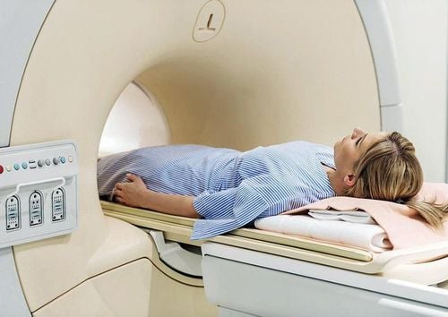
Chụp cộng hưởng từ ở Vinmec an toàn bậc nhất hiện nay bởi sự chính xác, không xâm lấn và không dùng tia X.
Vinmec International General Hospital put into use the magnetic resonance imaging machine 3.0 Tesla Silent technology. Magnetic resonance imaging machine 3.0 Tesla with Silent technology of GE Healthcare (USA).
Silent technology is especially beneficial for patients who are children, the elderly, weak health patients and patients undergoing surgery. Limiting noise, creating comfort and reducing stress for customers during the shooting process, helping to capture better quality images and shorten the shooting time. Magnetic resonance imaging technology is the technology applied in the most popular and safest imaging method today because of its accuracy, non-invasiveness and no use of X-rays. Customers can go directly to the system. Vinmec Health System nationwide to visit or contact the hotline HERE for support.
MORE:
MRI scan – the "golden" method to accurately diagnose disc herniation Bone scintigraphy detects early-stage bone metastatic cancer and other bone lesions First time using magnetic resonance imaging “no noise” at Vinmec Nha Trang