This is an automatically translated article.
The article was professionally consulted by Specialist Doctor I Tran Cong Trinh - Radiologist - Radiology Department - Vinmec Central Park International General Hospital.
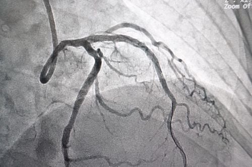
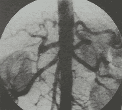
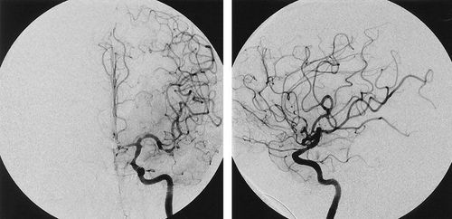
Digital subtraction angiography (DSA) and placement of a filter on the inferior vena cava to prevent large thrombus from moving into the pulmonary circulation and heart chambers, help patients reduce complications and mortality.
1. The role of digital imaging to erase the background and place the inferior vena cava filter
There are many causes of stenosis - obstruction of the superior and inferior vena cava systems (intravascular thrombosis, tumor compression...). Pulmonary embolism is one of the most dangerous complications in patients with venous thrombosis of the lower extremities or in the pelvis.Digital subtraction angiography (DSA) of veins and placement of inferior vena cava filters to prevent large thrombus from moving to the heart chambers and pulmonary circulation.
Based on the method of placement, there are 2 common procedures for mesh placement today:
Placement of the inferior vena cava through the right internal jugular vein Placement of the inferior vena cava through the common femoral vein
2. Indications and contraindications for background digitization and placement of inferior vena cava filter
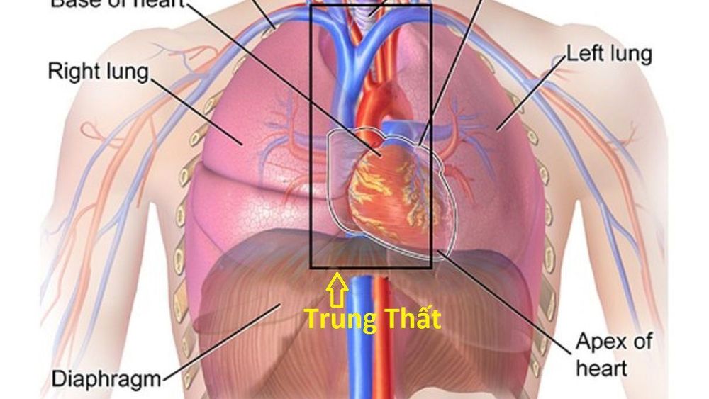
Chỉ định chụp DSA tĩnh mạch và đặt lưới lọc tĩnh mạch chủ dưới trong trường hợp u trung thất
Mediastinal tumor bronchial carcinoma causing vena cava obstruction Mediastinal lymphadenopathy due to metastatic cancer causing vena cava obstruction Lymphoma Hodgkin's or non-Hodgkin's causes vena cava to obstruct vena cava stenosis after radiotherapy and chemotherapy Venous occlusion due to fibrosis: After long-term catheterization (catheter), surgery, trauma, infection. Contraindicated and complicated with anticoagulation therapy Relatively indicated for large thrombus, suspended in the vascular lumen Contraindicated for venous DSA and inferior vena cava filter in case of:
Chronic thrombosis inferior vena cava Abnormalities in the anatomy of the vena cava impeding interventional techniques Severe renal failure (grade IV) Coagulopathy, loss of control. Pregnancy Reactions to iodinated contrast agents
3. Steps to conduct digital scan to remove background and place inferior vena cava filter
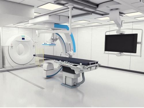
Máy chụp số hóa xóa nền
Before taking a job at Vinmec Central Park International General Hospital, the position of Doctor of Radiology from September 2017, Doctor Tran Cong Trinh worked at Gia Dinh People's Hospital since 2007. -2017. In his role, Dr. Tran Cong Trinh has participated in guiding the teaching of students, residents, specialists and new doctors entering the department
For examination and treatment at International General Hospital Vinmec, please come directly to Vinmec Health System or register online HERE.
SEE MORE
Application of digital erasure angiography (DSA) in cardiology In what cases is the electrocardiogram (electrocardiogram) used? How many types of coronary heart disease are there?


