This is an automatically translated article.
Posted by Resident Doctor, Master Tran Duc Tuan - Department of Diagnostic Imaging - Vinmec Central Park International General Hospital
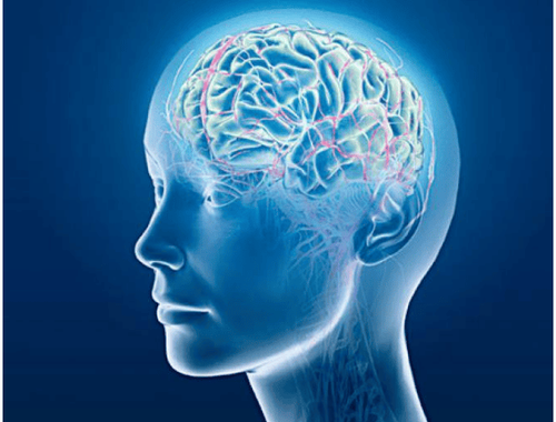
Thanks to the DSA background digitized angiogram system, many cardiovascular, cerebrovascular, and neurological diseases (especially myocardial infarction, stroke) have helped doctors in making a clear diagnosis and disease. timely intervention.
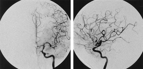
1. Digital angiography to erase the background in the neurological field
Digital Subtraction Angiography (DSA) is a new system of X-ray angiography. The purpose of this technique is to study blood vessels in the body and better see the lesions. and vascular disease before vascular intervention is indicated.Digital angiography with background erasure is an imaging method that combines X-ray and digital processing using algorithms to remove the background on 2 images obtained before and after contrast material is injected. into the patient's body, a full image of the heart and carotid, thoracic, abdominal and femoral arteries were obtained. This is the product of a combination of computerized image processing techniques and conventional angiography by Seldinger technique.
The basic principle of the digital background erasure angiography system is to use fluorescent light and X-ray to take pictures of the blood vessels at the sites to be checked before the contrast is injected and after the contrast is injected. into the blood vessel needed for imaging. Finally, to clarify the vascular system, the computer will erase the background image.
Components of a digital background removal angiography system typically include:
X-ray emitter Image acquisition unit Digital image processor: the central part of the system Display unit

Nguyên lý cơ bản của hệ thống chụp mạch số hóa xóa nền là dùng ánh sáng huỳnh quang và tia X chụp hình mạch máu ở những vị trí cần kiểm tra
First, the patient will be injected with a fluorescent dye called a contrast agent. This dye has the effect of brightening blood vessels, this dye is completely harmless and will be excreted from the body through the patient's urine. X-rays pass through the patient's body and are picked up by an Image Intensifier. A lens is placed between the bulb and the video camera for the purpose of limiting the intensity of light that reaches the camera. Then, the blood vessel to be imaged is injected with contrast material through a catheter that is inserted into the femoral artery through the skin. When the contrast material enters the body, the machine will acquire an animated image in a preset time unit. When there is no contrast agent as the background image, the image processing unit will take the acquired image (mask image) and proceed to exclude the background image with the image obtained when the contrast is present, which are static anatomical structures. similarity between the two images. Before the background digitized angiogram, the patient must fast from breakfast and be well prepared, including tests such as:
Record electrocardiogram Blood test Chest X-ray technique Digital angiogram remove background It is an invasive imaging technique and carries a risk of complications when taking pictures. Therefore, it must be strictly prescribed and must be agreed to by the patient. After the scan, the patient will be bandaged in the groin area and lying motionless for 24 hours, and absolutely avoiding leg movement.
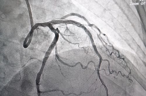
2. Applications of DSA

Chụp DSA để làm gì?
Evaluation of anomalies of the carotid artery, aorta, renal artery, limb artery and peripheral arteries . Cardiac catheterization, closure of the cardiac catheterization, angioplasty, and intra-aortic balloon counterpulsation. Place a vena cava filter, place a pacemaker, remove a foreign body from the circulatory system, and echocardiographically and intracardiacally. Exploration and treatment of electrophysiology, uterine tumor or liver cancer, brain tumor, and cerebrovascular abnormalities... Digital angiography technology has helped doctors to detect conditions early. Abnormalities of the blood flow, help in the accurate diagnosis and treatment of serious diseases in the neurological field, plan the anatomy and help to accurately determine the location of the damage inside the body.
Thanks to this DSA machine, quite a few cases of neurological diseases, especially cerebrovascular diseases, were clearly diagnosed and were quickly and promptly intervened within the golden period, so their lives were saved.
Please dial HOTLINE for more information or register for an appointment HERE. Download MyVinmec app to make appointments faster and to manage your bookings easily.