This is an automatically translated article.
The article was professionally consulted by Specialist Doctor I Tran Cong Trinh - Radiologist - Radiology Department - Vinmec Central Park International General Hospital. The doctor has many years of experience in the field of diagnostic imaging.1. Doppler ultrasound
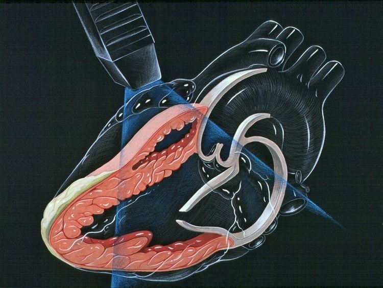
Doppler ultrasound is medical ultrasound that uses the Doppler effect to produce moving images of body tissues and fluids and their relative velocities to the transmitter. Doppler ultrasound is a type of ultrasound with high accuracy and safety, divided into 2 types:Continuous Doppler: An ultrasound consisting of 2 crystals in a transducer with 2 different functions. In it, one crystal has the function of emitting sound waves and the other crystal is responsible for sound waves. From there, continuous Doppler ultrasound can check at high velocities. However, the form cannot check the location of the return point. Pulsed Doppler: An ultrasound with only one crystal at the tip. That means pulsed Doppler ultrasound can both transmit and receive sound waves. At the transducer part of the pulse Doppler ultrasound machine, there is a connection of longitudinal pulse chains for the purpose of emitting sound waves in the scanning direction of each transducer.

2. Principle of Doppler ultrasound
Doppler data is displayed graphically using spectral Doppler or as images using color or directional Doppler or power Doppler also known as non-directional Doppler. This Doppler shift is in the audible range and is often clearly presented using stereo speakers: this produces a very distinctive, albeit synthetic, sound like a beat.All modern ultrasound scanners use pulsed Doppler to measure velocity. The pulse wave device transmits and receives a sequence of pulses. The frequency change of each pulse is ignored, however the relative phase change of the pulses is used to obtain the frequency change, since frequency is the rate of change of phase. The main advantage of pulse wave Doppler over continuous wave is that the distance information is obtained, the time between transmitted and received pulses multiplied by the speed of sound equal to the distance, and gain correction is applied. The disadvantage of pulsed Doppler is that the measurements can be aliased. The terms Doppler ultrasound have been accepted to apply to both pulsed and continuous Doppler systems, although they use different mechanisms for measuring velocity.
3. Doppler ultrasound technique
3.1 Carotid Doppler Ultrasound Performed: With the patient lying on his back, the ultrasound probe is scanned over the neck area to display an ultrasound image of the blood vessels in the neck (including the carotid vessels) on the computer screen.

Using Doppler ultrasound technique is a way to help detect and diagnose some diseases quickly and effectively. If you suspect you have a problem, you can come to Vinmec International General Hospital System, one of the leading prestigious hospitals in the country, Vinmec uses current generations of color ultrasound machines. the most modern in examination and treatment for patients. One of them is GE Healthcarecar's Logig E9 ultrasound machine with full options, HD resolution probes for clear images, accurate assessment of lesions. In addition, a team of experienced doctors and nurses will greatly assist in the diagnosis and early detection of abnormal signs of the body in order to provide timely treatment.
Please dial HOTLINE for more information or register for an appointment HERE. Download MyVinmec app to make appointments faster and to manage your bookings easily.


